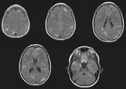
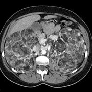
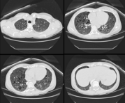
This is a set of images from an MRI of the brain in a patient with TSC.This is an image from a contrast-enhanced CT of the abdomen in another patient with TSC.This computed tomography image shows randomly arranged cysts in both lungs.  The patient had TSC and a renal AML.
Find information on thousands of medical conditions and prescription drugs.

Tuberous Sclerosis

Tuberous sclerosis, (meaning "hard potatoes"), is a rare genetic disorder primarily characterized by a triad of seizures, mental retardation, and skin lesions (called adenoma sebaceum). This "classic" Vogt triad is present in 30-50% of cases; in particular, up to 30% of tuberous sclerosis reportedly have normal mentation. Tuberous sclerosis, along with Neurofibromatosis type I, Neurofibromatosis type II (a.k.a. MISME syndrome), Sturge-Weber, and Von Hippel-Lindau compromise the Phakomatoses or neurocutaneous syndromes, all of which have neurologic and dermatologic lesions. more...

Home
Diseases
A
B
C
D
E
F
G
H
I
J
K
L
M
N
O
P
Q
R
S
T
Candidiasis
Tachycardia
Taeniasis
Talipes equinovarus
TAR syndrome
Tardive dyskinesia
Tarsal tunnel syndrome
Tay syndrome ichthyosis
Tay-Sachs disease
Telangiectasia
Telangiectasia,...
TEN
Teratoma
Teratophobia
Testotoxicosis
Tetanus
Tetraploidy
Thalassemia
Thalassemia major
Thalassemia minor
Thalassophobia
Thanatophobia
Thoracic outlet syndrome
Thrombocytopenia
Thrombocytosis
Thrombotic...
Thymoma
Thyroid cancer
Tick paralysis
Tick-borne encephalitis
Tietz syndrome
Tinnitus
Todd's paralysis
Topophobia
Torticollis
Touraine-Solente-Golé...
Tourette syndrome
Toxic shock syndrome
Toxocariasis
Toxoplasmosis
Tracheoesophageal fistula
Trachoma
Transient...
Transient Global Amnesia
Transposition of great...
Transverse myelitis
Traumatophobia
Treacher Collins syndrome
Tremor hereditary essential
Trichinellosis
Trichinosis
Trichomoniasis
Trichotillomania
Tricuspid atresia
Trigeminal neuralgia
Trigger thumb
Trimethylaminuria
Triplo X Syndrome
Triploidy
Trisomy
Tropical sprue
Tropophobia
Trypanophobia
Tuberculosis
Tuberous Sclerosis
Tularemia
Tungiasis
Turcot syndrome
Turner's syndrome
Typhoid
Typhus
Tyrosinemia
U
V
W
X
Y
Z
Medicines

This grouping is an artifact of an earlier time in medicine, before the distinct genetic basis of each of these diseases was understood.

The neuropathologic findings of the cortical "tubers" (sclerose tubereuse) was first described by Désiré-Magloire Bourneville in 1880.

Individuals with tuberous sclerosis may experience none or all of the symptoms with varying degrees of severity. Tuberous sclerosis is a multi-system disease that can affect the brain, kidneys, heart, eyes, lungs, and other organs. Small benign tumors may grow on the face and eyes, as well as in the brain, kidneys, and other organs. Neuroimaging studies may be able to confirm the diagnosis. Seizures most often begin in the first year of life.

Tuberous sclerosis' acronym is T.S.C. (Tuberous sclerosis complex) so as to avoid confusion with Tourette's Syndrome.

Genetics

Tuberous sclerosis (TSC) is a genetic disorder caused by mutations on either of two genes TSC1 and TSC2. It has an autosomal dominant pattern of inheritance and penetrance is 100%. The incidence is between 1/6,000 and 1/10,000. One third of cases are inherited; the rest are new mutations.

TSC1 is located on chromosome 9q34 and encodes for the protein hamartin. It was discovered in 1997. TSC2 is located on chromosome 16p13.3 and encodes for the protein tuberin. It was discovered in 1993. TSC2 is contiguous with PKD1 (which causes one form of polycystic kidney disease). Gross deletions affecting both genes may account for the 2% of individuals with TSC who develop PKD in childhood.

TSC1 and TSC2 are both tumor suppressor genes that function according to Knudson's "two hit" hypothesis. That is, a second random mutation must occur before a tumor can develop. This explains the wide expressivity of the disease. Of the two, TSC2 has been associated with a more severe form of TSC. However, the difference is subtle and statistical and cannot be used to identify the mutation clinically. Estimates of the proportion of TSC caused by TSC2 ranges from 55% to 80-90%. Current genetic tests have difficulty locating the mutation in approximately 20% of individuals.

Hamartin/tuberin function as a complex, whose biological activity appears to be a Rheb GTPase. They function within the growth factor (insulin) signalling pathway and are involved in suppressing mTOR signalling.

Read more at Wikipedia.org


[List your site here Free!]


Pulmonary cysts consistent with lymphangioleiomyomatosis are common in women with tuberous sclerosis ; genetic and radiographic analysis
From CHEST, 3/1/02 by Francis McCormack

Abbreviations: LAM = lymphangioleiomyomatosis; TSC = tuberous sclerosis

Lymphangioleiomyomatosis (LAM) and multifocal micronodular pneumocyte hyperplasia produce cystic and nodular disease, respectively, in the lungs of patients with tuberous sclerosis (TSC). The objective of this study was to prospectively characterize the prevalence, clinical presentation, and genetic basis of lung disease in TSC. CT scanning of the chest on 23 asymptomatic women with TSC identified cystic or nodular changes in 52%. Cystic pulmonary parenchymal changes consistent with LAM were found in nine patients (39%). These patients tended to be older than cyst-negative patients (31.9 [+ or -] 7.6 years vs 24.8 [+ or -] 11.6 years, p = 0.09), and there was no correlation between presence of cysts and tobacco use, age at menarche, history of pregnancy, or use of estrogen-containing medications. Three of the cyst-positive patients had a prior history of pneumothorax. Pulmonary function studies revealed evidence of gas trapping but normal spirometric indexes in the cyst-positive group. All 9 cyst-positive patients had angiomyolipomas, which were larger (p < 0.05) and more frequently required intervention (p = 0.08) than cyst-negative patients (8 of 14 patients with angiomyolipomas, p < 0.05). Ten patients (43%) had pulmonary parenchymal nodules. Pulmonary nodules were more common in women with cysts (78% vs 21%, p < 0.05). TSC2 mutations were identified in all cyst-positive patients who were tested (n = 8), while both TSC1 and TSC2 mutations were identified in patients with nodular disease. Correlation of the mutational and radiographic data revealed one pair of sisters who were discordant for cystic disease, two mother-daughter pairs who were discordant for nodular disease, and no clear association between cyst development and a specific mutational type. This prospective analysis demonstrates that cystic and nodular pulmonary changes consistent with LAM and multifocal modular pneumocyte hyperplasia are common in women with TSC.

* From the Departments of Pediatrics (Dr. Sethuraman), Neurology (Dr. Franz, Ms. Leonard, Ms. Chuck), Radiology (Drs. Brody and Meyer), and Medicine (Dr. McCormack), Division of Pulmonary and Critical Care Medicine, University of Cincinnati, Cincinnati, OH; the Department of Pathology (Dr. Colby), Mayo Clinic, Scottsdale, AZ; and the Department of Medicine, Division of Hematology (Drs. Dabora and Kwiatkowski), Brigham and Women's Hospital, Boston, MA.

Correspondence to: Frank McCormack, MD, Division of Pulmonary and Critical Care Medicine, MSB Room 6001, 231 Albert Sabin Way, Cincinnati, OH 45267-0564; e-mail:frank.mccormack@uc.edu

COPYRIGHT 2002 American College of Chest Physicians
COPYRIGHT 2002 Gale Group

Return to Tuberous Sclerosis
Home Contact Resources Exchange Links ebay