Hookworm life cycleCivilian Public Service workers built and installed 2065 privies for hookworm eradication in Mississippi and Florida from 1943 to 1947.
Find information on thousands of medical conditions and prescription drugs.

Ankylostomiasis

The hookworm is a parasite that lives in the small intestine of its host, which may be a mammal such as a dog, cat, or human. Two species of hookworms commonly infect humans, Ancylostoma duodenale and Necator americanus. The distribution of each species significantly overlaps that of the other. Necator americanus predominates in The Americas, Sub-Saharan Africa, Southeast Asia, China and Indonesia, while A. duodenale predominates in the Middle East, North Africa, India and (formerly) in southern Europe. more...

Home
Diseases
A
Aagenaes syndrome
Aarskog Ose Pande syndrome
Aarskog syndrome
Aase Smith syndrome
Aase syndrome
ABCD syndrome
Abdallat Davis Farrage...
Abdominal aortic aneurysm
Abdominal cystic...
Abdominal defects
Ablutophobia
Absence of Gluteal muscle
Acalvaria
Acanthocheilonemiasis
Acanthocytosis
Acarophobia
Acatalasemia
Accessory pancreas
Achalasia
Achard syndrome
Achard-Thiers syndrome
Acheiropodia
Achondrogenesis
Achondrogenesis type 1A
Achondrogenesis type 1B
Achondroplasia
Achondroplastic dwarfism
Achromatopsia
Acid maltase deficiency
Ackerman syndrome
Acne
Acne rosacea
Acoustic neuroma
Acquired ichthyosis
Acquired syphilis
Acrofacial dysostosis,...
Acromegaly
Acrophobia
Acrospiroma
Actinomycosis
Activated protein C...
Acute febrile...
Acute intermittent porphyria
Acute lymphoblastic leukemia
Acute lymphocytic leukemia
Acute mountain sickness
Acute myelocytic leukemia
Acute myelogenous leukemia
Acute necrotizing...
Acute promyelocytic leukemia
Acute renal failure
Acute respiratory...
Acute tubular necrosis
Adams Nance syndrome
Adams-Oliver syndrome
Addison's disease
Adducted thumb syndrome...
Adenoid cystic carcinoma
Adenoma
Adenomyosis
Adenosine deaminase...
Adenosine monophosphate...
Adie syndrome
Adrenal incidentaloma
Adrenal insufficiency
Adrenocortical carcinoma
Adrenogenital syndrome
Adrenoleukodystrophy
Aerophobia
Agoraphobia
Agrizoophobia
Agyrophobia
Aicardi syndrome
Aichmophobia
AIDS
AIDS Dementia Complex
Ainhum
Albinism
Albright's hereditary...
Albuminurophobia
Alcaptonuria
Alcohol fetopathy
Alcoholic hepatitis
Alcoholic liver cirrhosis
Alektorophobia
Alexander disease
Alien hand syndrome
Alkaptonuria
Alliumphobia
Alopecia
Alopecia areata
Alopecia totalis
Alopecia universalis
Alpers disease
Alpha 1-antitrypsin...
Alpha-mannosidosis
Alport syndrome
Alternating hemiplegia
Alzheimer's disease
Amaurosis
Amblyopia
Ambras syndrome
Amelogenesis imperfecta
Amenorrhea
American trypanosomiasis
Amoebiasis
Amyloidosis
Amyotrophic lateral...
Anaphylaxis
Androgen insensitivity...
Anemia
Anemia, Diamond-Blackfan
Anemia, Pernicious
Anemia, Sideroblastic
Anemophobia
Anencephaly
Aneurysm
Aneurysm
Aneurysm of sinus of...
Angelman syndrome
Anguillulosis
Aniridia
Anisakiasis
Ankylosing spondylitis
Ankylostomiasis
Annular pancreas
Anorchidism
Anorexia nervosa
Anosmia
Anotia
Anthophobia
Anthrax disease
Antiphospholipid syndrome
Antisocial personality...
Antithrombin deficiency,...
Anton's syndrome
Aortic aneurysm
Aortic coarctation
Aortic dissection
Aortic valve stenosis
Apert syndrome
Aphthous stomatitis
Apiphobia
Aplastic anemia
Appendicitis
Apraxia
Arachnoiditis
Argininosuccinate...
Argininosuccinic aciduria
Argyria
Arnold-Chiari malformation
Arrhythmogenic right...
Arteriovenous malformation
Arteritis
Arthritis
Arthritis, Juvenile
Arthrogryposis
Arthrogryposis multiplex...
Asbestosis
Ascariasis
Aseptic meningitis
Asherman's syndrome
Aspartylglycosaminuria
Aspergillosis
Asphyxia neonatorum
Asthenia
Asthenia
Asthenophobia
Asthma
Astrocytoma
Ataxia telangiectasia
Atelectasis
Atelosteogenesis, type II
Atherosclerosis
Athetosis
Atopic Dermatitis
Atrial septal defect
Atrioventricular septal...
Atrophy
Attention Deficit...
Autoimmune hepatitis
Autoimmune...
Automysophobia
Autonomic dysfunction
Familial Alzheimer disease
Senescence
B
C
D
E
F
G
H
I
J
K
L
M
N
O
P
Q
R
S
T
U
V
W
X
Y
Z
Medicines

Hookworms are thought to infect 800 million people worldwide. The A. braziliense and A. tubaeforme species infect cats, while A. caninum infects dogs. Uncinaria stenocephala infects both dogs and cats.

These worms are much smaller than the large roundworm, Ascaris lumbricoides, and the complications of tissue migration and mechanical obstruction so frequently observed with roundworm infestation are less frequent in hookworm infestation. The most significant risk of hookworm infection is anemia secondary to loss of iron (and protein) into the gut.

Ankylostomiasis, alternatively spelled anchylostomiasis and also called helminthiasis, "miners' anaemia", and in Germany Wurmkrankheit is the disease caused by hookworms.

It was prevalent in the mining industry in England, France, Germany, Belgium, North Queensland and elsewhere. It was known in Egypt even in very ancient times.

It caused a great mortality among blacks in the West Indies towards the end of the 18th century; and through descriptions sent from Brazil and various other tropical and sub-tropical regions.

The disease was linked to nematoid worms (Ankylostoma duodenalis) from one-third to half an inch long in the intestine chiefly through the labours of Bilharz and Griesinger in Egypt (1854).

The symptoms, as first observed among blacks, were pain in the stomach, capricious appetite, pica (or dirt-eating), obstinate constipation followed by diarrhoea, palpitations, small and unsteady pulse, coldness of the skin, pallor of the skin and mucous membranes, diminution of the secretions, loss of strength and, in cases running a fatal course, dysentery, haemorrhages and dropsies.

The disease was first known in Europe among the Italian workmen employed on the St Gotthard tunnel. In contrast with most intestinal helminthiases that concentrate parasitic load in children, hookworm prevalence is often higher among adult males. In tropical areas this is associated with high prevalence of anemia among adult men.

Hookworms are leading causes of maternal and child morbidity in the developing countries of the tropics and subtropics. In susceptible children hookworms cause intellectual, cognitive and growth retardation; as well as intrauterine growth retardation, prematurity and low birth weight among newborns born to infected mothers. Hookworm infection is rarely fatal, but anemia can be significant in the heavily infected individual.

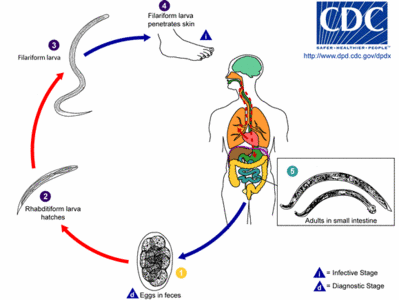
Hookworm life cycle

See the image for the biological life cycle of the hookworm.

The hookworm larva lives in infected soil. The larva penetrates the skin on contact, e.g. when walking barefoot, enters the bloodstream and is transported to the lungs. From the lungs and bronchi it transfers to the stomach and intestine by coughing up and swallowing phlegm. The larva clings to the intestinal mucous membrane and develops into an adult hookworm, drawing its nourishment from the blood-vessels of their host, and as the parasites are found in hundreds in the body after death, the disorders of digestion, the increasing anaemia and the consequent dropsies and other cachectic symptoms are easily explained. The adult releases eggs that leave the human body with the feces. The eggs hatch in soil and eventually develop into larva.

Read more at Wikipedia.org


[List your site here Free!]


Eosinophilic pleural effusion due to artemisnin: a case report
From CHEST, 10/1/05 by Mayank Vats

INTRODUCTION: Drug induced eosinophilic pleural effusion (EPE) is well documented in literature. EPE is not a disease rather an interesting laboratory finding, defined as >10% eosinophils in pleural fluid, exclusive of erythrocytes. Pleural fluid eosinophilia may be associated with blood eosinophilia e.g. Loeffler syndrome, Churg Strauss syndrome etc. Conversly EPE can also occur without blood eosinophilia e.g. Pulmonary Infarction, Pneumonia or Trauma. We report a case of bilateral EPE secondary to Artemisnin with no peripheral eosinophilia.

CASE PRESENTATION: AM 46-year-old female presented to OPD because of bilateral dull aching chest pain, which increased, on taking deep breath. Five days before the presenting illness she also had high grade fever with chills for which she received injection Artemisinin on the suspicion of malaria in another tertiary health care center. The patient demonstrated no symptom, sign or laboratory data of any infectious process, Chest X-Ray PA revealed bilateral pleural effusion more on right side & no parenchymal infiltrates. Patient underwent right thoracentesis, revealing protein-4.2 g/dl, 60%-lymphocyte, 26%-eosinophils, 10%-mesothelial cells & 4%-polymorphs. Next day, left thoracentesis revealed protein-4.4 g/dl, 56%-lymphocytes, 32%-eosinophils, 8%-mesothelial cell & 4%-polymorphs. Pleural biopsy showed mixed lymphocytic & eosinophilic infiltrates within pleura. The bacterial, fungal & mycobacterial smear & culture of fluid & biopsy specimen were negative after 6 weeks. Her serology was negative for Anti-Nuclear Antibody, LE cells, & Rheumatoid factor. There was no history of contact of pulmonary tuberculosis & Montoux test was negative. A detailed history of illness (including absence of any significant past medical history), drug intake & complete evaluation strongly pointed out towards a possible drug (Artemisnin) induced pleural effusion, hence after informed consent & with permission of ethical society of institution, injection Artemisnin 40mg IM x 5 days was given, as challenge dose & on 7th day patient again developed bilateral chest pain & Chest X-Ray revealed bilateral minimal pleural effusion. Patient was started on prednisolone 60 mg/day for 7 Days & pleura] effusion disappeared completely, hence confirming the diagnosis.

DISCUSSIONS: Air & Blood are the most common cause of EPE. Other causes of EPE1 are Bronchial or Pleural malignancy, hypersensitivity reactions, pulmonary infarction & infection with viruses, fungus (2) (Coccidiodomycosis, Histoplasmosis) & parasites (Echinococcus, Amoebiasis, Ascariaris, Schistosomiasis, Ankylostomiasis etc.). Drug reactions like Dantrolene, Bromocriptine, Nitrofurantoin. Procarbazine, Ergot, Methotrexate has been implicated in EPE. Allergic disease like Asthma, Tropical Pulmonary Eosonipbilia, Churg-Strauss syndrome may also lead to Eosinophilic Pleural Effusion. (3) In this case the absence of other causes for the EPE & its complete resolution after withdrawal of Artemisnin, reappearance on challenging with low dose supports a causative relationship of this drug with the development of EPE. This is probably the first case report of EPE secondary to Artemisnin.

CONCLUSION: Artemisnin must be included in the list of drugs causing EPE, however the exact mechanism is not known, but, if the drug is considered as a cause of EPE, the drug should be immediately discontinued. Clinical Implications: a high degree of suspicion should always be kept in mind to prove the drug as an etiologic agent in undiagnosed EPE, and a challenge test with all due precautions should always be done to prove or disprove this etiology.

REFERENCES:

(1) Campbell GD, Webb WR: Eosinophilic Pleural Effusion, Amer. Rev. Resp. Dis. 1964, 90, 194.

(2) Curran WS, Williams AW: Eosinophilic Pleural Effusion, Arch. Inern. Med. (Chiacgo) 1963, 111, 809.

(3) Erzurum SE, Underwood GA, Hamilos DL, Waldron JA: Pleural eff

DISCLOSURE: Mayank Vats, None.

Mayank Vats MD * Rakesh C. Gupta MD Deepa V. Khandelwal MBBS Manohar L. Gupta MD Neeraj Gupta MD Mukesh Tailor MBBS J.L.N. Medical College, Ajmer, Rajasthan, India

COPYRIGHT 2005 American College of Chest Physicians
COPYRIGHT 2005 Gale Group

Return to Ankylostomiasis
Home Contact Resources Exchange Links ebay