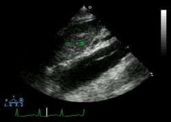
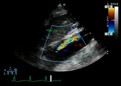
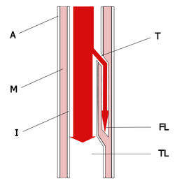
Illustration of an aortic dissectionEchocardiogram of an aortic dissectionEchocardiogram of an aortic dissection
Find information on thousands of medical conditions and prescription drugs.

Aortic dissection

Aortic dissection is a tear in the wall of the aorta (the largest artery of the body). This tear causes blood to flow between the layers of the wall of the aorta and dissects the layers apart. Aortic dissection is a medical emergency and can quickly lead to death, even with optimal treatment. more...

Home
Diseases
A
Aagenaes syndrome
Aarskog Ose Pande syndrome
Aarskog syndrome
Aase Smith syndrome
Aase syndrome
ABCD syndrome
Abdallat Davis Farrage...
Abdominal aortic aneurysm
Abdominal cystic...
Abdominal defects
Ablutophobia
Absence of Gluteal muscle
Acalvaria
Acanthocheilonemiasis
Acanthocytosis
Acarophobia
Acatalasemia
Accessory pancreas
Achalasia
Achard syndrome
Achard-Thiers syndrome
Acheiropodia
Achondrogenesis
Achondrogenesis type 1A
Achondrogenesis type 1B
Achondroplasia
Achondroplastic dwarfism
Achromatopsia
Acid maltase deficiency
Ackerman syndrome
Acne
Acne rosacea
Acoustic neuroma
Acquired ichthyosis
Acquired syphilis
Acrofacial dysostosis,...
Acromegaly
Acrophobia
Acrospiroma
Actinomycosis
Activated protein C...
Acute febrile...
Acute intermittent porphyria
Acute lymphoblastic leukemia
Acute lymphocytic leukemia
Acute mountain sickness
Acute myelocytic leukemia
Acute myelogenous leukemia
Acute necrotizing...
Acute promyelocytic leukemia
Acute renal failure
Acute respiratory...
Acute tubular necrosis
Adams Nance syndrome
Adams-Oliver syndrome
Addison's disease
Adducted thumb syndrome...
Adenoid cystic carcinoma
Adenoma
Adenomyosis
Adenosine deaminase...
Adenosine monophosphate...
Adie syndrome
Adrenal incidentaloma
Adrenal insufficiency
Adrenocortical carcinoma
Adrenogenital syndrome
Adrenoleukodystrophy
Aerophobia
Agoraphobia
Agrizoophobia
Agyrophobia
Aicardi syndrome
Aichmophobia
AIDS
AIDS Dementia Complex
Ainhum
Albinism
Albright's hereditary...
Albuminurophobia
Alcaptonuria
Alcohol fetopathy
Alcoholic hepatitis
Alcoholic liver cirrhosis
Alektorophobia
Alexander disease
Alien hand syndrome
Alkaptonuria
Alliumphobia
Alopecia
Alopecia areata
Alopecia totalis
Alopecia universalis
Alpers disease
Alpha 1-antitrypsin...
Alpha-mannosidosis
Alport syndrome
Alternating hemiplegia
Alzheimer's disease
Amaurosis
Amblyopia
Ambras syndrome
Amelogenesis imperfecta
Amenorrhea
American trypanosomiasis
Amoebiasis
Amyloidosis
Amyotrophic lateral...
Anaphylaxis
Androgen insensitivity...
Anemia
Anemia, Diamond-Blackfan
Anemia, Pernicious
Anemia, Sideroblastic
Anemophobia
Anencephaly
Aneurysm
Aneurysm
Aneurysm of sinus of...
Angelman syndrome
Anguillulosis
Aniridia
Anisakiasis
Ankylosing spondylitis
Ankylostomiasis
Annular pancreas
Anorchidism
Anorexia nervosa
Anosmia
Anotia
Anthophobia
Anthrax disease
Antiphospholipid syndrome
Antisocial personality...
Antithrombin deficiency,...
Anton's syndrome
Aortic aneurysm
Aortic coarctation
Aortic dissection
Aortic valve stenosis
Apert syndrome
Aphthous stomatitis
Apiphobia
Aplastic anemia
Appendicitis
Apraxia
Arachnoiditis
Argininosuccinate...
Argininosuccinic aciduria
Argyria
Arnold-Chiari malformation
Arrhythmogenic right...
Arteriovenous malformation
Arteritis
Arthritis
Arthritis, Juvenile
Arthrogryposis
Arthrogryposis multiplex...
Asbestosis
Ascariasis
Aseptic meningitis
Asherman's syndrome
Aspartylglycosaminuria
Aspergillosis
Asphyxia neonatorum
Asthenia
Asthenia
Asthenophobia
Asthma
Astrocytoma
Ataxia telangiectasia
Atelectasis
Atelosteogenesis, type II
Atherosclerosis
Athetosis
Atopic Dermatitis
Atrial septal defect
Atrioventricular septal...
Atrophy
Attention Deficit...
Autoimmune hepatitis
Autoimmune...
Automysophobia
Autonomic dysfunction
Familial Alzheimer disease
Senescence
B
C
D
E
F
G
H
I
J
K
L
M
N
O
P
Q
R
S
T
U
V
W
X
Y
Z
Medicines

Overview

As with all other arteries, the aorta is made up of three layers. The layer that is in direct contact with the flow of blood is the tunica intima, commonly called the intima. This layer is made up of mainly endothelial cells. Just deep to this layer is the tunica media, known as the media. This "middle layer" is made up of smooth muscle cells and elastic tissue. The outermost layer (furthest from the flow of blood) is known as the tunica adventitia or the adventitia. This layer is composed of connective tissue.

In an aortic dissection, blood penetrates the intima and enters the media layer. The high pressure rips the tissue of the media apart, allowing more blood to enter. This can propagate along the length of the aorta for a variable distance, dissecting either towards or away from the heart or both. The initial tear is usually within 10 cm of the aortic valve.

The risk in aortic dissection is that the aorta may rupture, leading to massive blood loss resulting in death.

Classification systems

Several different classification systems have been used to describe aortic dissections. The systems commonly in use are either based on the anatomy of the dissection or the duration of onset of symptoms prior to presentation.

DeBakey classification system

The DeBakey system is an anatomical description of the aortic dissection. It categorizes the dissection based on where the original intimal tear is located and the extent of the dissection (localized to either the ascending aorta or descending aorta, or involves both the ascending and descending aorta.

  • Type I - Originates in ascending aorta, propagates at least to the aortic arch and often beyond it distally.
  • Type II – Originates in and is confined to the ascending aorta.
  • Type III – Originates in descending aorta, rarely extends proximally.

Pathophysiology

The initiating event in an aortic dissection is a tear in the intimal lining of the aorta. Due to the high pressures in the aorta, blood enters the media at the point of the tear. The force of the blood entering the media causes the tear to extend. It may extend proximally (closer to the heart) or distally (away from the heart) or both. The blood will travel through the media, creating a false lumen (the true lumen is the normal conduit of blood in the aorta). Separating the false lumen from the true lumen is a layer of intimal tissue. This tissue is known as the intimal flap.

The vast majority of aortic dissections originate with an intimal tear in either the ascending aorta (65%), the aortic arch (10%), or just distal to the ligamentum arteriosum in the descending thoracic aorta (20%).

Read more at Wikipedia.org


[List your site here Free!]


Aortic Dissection
From Archives of Pathology & Laboratory Medicine, 5/1/04 by Xie, Linjun

A 64-year-old white woman with a past medical history of osteoarthritis, osteoporosis, hyperthyroidism, and hypercholesterolemia presented with chest and neck pain, headache, blurring of vision, and numbness of the right lower extremity for a few hours. She was found to have a pulse rate of 60 beats per minute, respiratory rate of 18 breaths per minute, and blood pressure of 98 mm Hg, palpable. Heart and lung sounds were unremarkable. Her right leg was pale, cold, and numb with decreased touch and pain sensation. Her laboratory data showed a creatine kinase level of 414 U/L (reference range, 260-140 U/L), creatine kinase MB fraction of 17.5 U/L (reference range, 0-13.9 U/L), and troponin level less than 0.01 × 10^sup -6^ mg/ mL. Electrocardiogram revealed sinus rhythm at 60 beats per minute and left bundle branch block. Chest radiography showed normal lung fields without mediastinal widening. Doppler study of the lower extremities found no flow in the right leg and normal flow in the left leg. Head computed tomography was negative for hemorrhage or tumor. Her condition progressively deteriorated, and the patient died 1 day after admission. A complete autopsy was performed.

The pericardial sac was found to be distended with the formation of hemopericardium (400 mL of blood and blood clots). The ascending aorta was enlarged and distended (Figure 1). The outer surface was smooth, glistening, and dark brown. On opening, the aorta showed dissection involving ascending and descending portions (Figure 1, inset; Figure 2). The false lumen between the intima and adventitia was filled with a large amount of blood and blood clots (Figure 1, inset; Figure 3). There was a tear in the intima in the right lateral wall of the ascending aorta where usually the greatest shear force on the artery wall results from blood expelled from the heart under high pressure. The entry of blood between the intima and adventitia extended forward and all the way down the descending aorta, terminating just at the junction between the descending aorta and the iliac artery (Figure 2). The dissection also extended proximally toward the heart. Passage of blood into the pericardial cavity was retrograde through a small channel communicating with the false lumen. Acute onset of hemopericardium resulted in cardiac tamponade, which was the main cause of death for this patient. Microscopically, the dissecting hematoma spread along the laminar planes of the media between the middle and outer thirds, and elastic staining of an aortic wall specimen did not demonstrate cystic medial degeneration (Figure 3). Other significant findings included hemorrhage in the mediastinum and the right ureter, left ventricular hypertrophy, and mild atherosclerosis of the left circumflex, right coronary, and basal arteries.

In elderly patients, hypertension is the most common cause of aortic dissection.1 However, this patient had no previous history of hypertension and clinically manifested with hypotension rather than hypertension. Recent clinical investigations of 274 cases demonstrated that 49% of patients with aortic dissection have elevated blood pressure, 31% have pulse deficits, and 17% have focal neurological deficits. The presence of pulse deficits or focal neurological deficits increases the likelihood of an acute aortic dissection.2 Systemic or localized abnormalities of connective tissue that affect the aorta, such as Marfan syndrome, should be investigated. The most frequent preexisting, histologically detectable lesion is cystic medial degeneration, which consists of elastic tissue fragmentation and separation of the elastic and fibromuscular elements of the tunica media by small cystic spaces filled with amorphous extracellular matrix.1 The elastic stain is advantageous for the detection of cystic medial degeneration.

References

1. Cotran RS, Kumar V, Collins T. Robbins Pathologic Basis of Disease. 6th ed. Philadelphia, Pa: WB Saunders Co; 1999:526-529.

2. Klompas M. Does this patient have an acute thoracic aortic dissection? JAMA. 2002;287:2262-2271.

Linjun Xie, MD; Henry J. Shih, MD; Lester Freedman, MD

Accepted for publication January 7, 2004.

From the Department of Pathology, Nassau University Medical Center, East Meadow, NY.

The authors have no relevant financial interest in the products or companies described in this article.

Reprints: Linjun Xie, MD, Department of Pathology, Box 47, Nassau University Medical Center, 2201 Hempstead Turnpike, East Meadow, NY 11554 (e-mail: lxie555@hotmail.com).

Copyright College of American Pathologists May 2004
Provided by ProQuest Information and Learning Company. All rights Reserved

Return to Aortic dissection
Home Contact Resources Exchange Links ebay