Aortic valve stenosis
Aortic valve stenosis (AS) is a heart condition caused by the incomplete opening of the aortic valve. more...
The aortic valve controls the direction of blood flow from the left ventricle to the aorta. When in good working order, the aortic valve does not impede the flow of blood between these two spaces. Under some circumstances, the aortic valve becomes narrower than normal, impeding the flow of blood. This is known as aortic valve stenosis, or aortic stenosis, often abbreviated as AS.
Pathophysiology
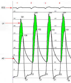
When the aortic valve becomes stenotic, it causes a pressure gradient between the left ventricle (LV) and the aorta. The more constricted the valve, the higher the gradient between the LV and the aorta. For instance, with a mild AS, the gradient may be 20 mmHg. This means that, at peak systole, while the LV may generate a pressure of 140 mmHg, the pressure that is transmitted to the aorta will only be 120 mmHg. So, while a blood pressure cuff may measure a normal systolic blood pressure, the actual pressure generated by the LV would be considerably higher.
In individuals with AS, the left ventricle (LV) has to generate an increased pressure in order to overcome the increased afterload caused by the stenotic aortic valve and eject blood out of the LV. The more severe the aortic stenosis, the higher the gradient is between the left ventricular systolic pressures and the aortic systolic pressures. Due to the increased pressures generated by the left ventricle, the myocardium (muscle) of the LV undergoes hypertrophy (increase in muscle mass). This is seen as thickening of the walls of the LV. The type of hypertrophy most commonly seen in AS is concentric hypertrophy, meaning that all the walls of the LV are (approximately) equally thickened.
Etiology
Causes of aortic stenosis include acute rheumatic fever, bicuspid aortic valve and congenital anomalies. As individuals age, calcification of the aortic valves may occur and result in stenosis.
Physical examination
It is most often diagnosed when it is asymptomatic. It is found on routine examination of the heart. A fairly loud systolic, crescendo-decrescendo murmur is heard loudest at the upper right sternal border, and radiates to the carotid arteries. The murmur increases with squatting, decreases with standing and isometric muscular contraction, which helps distinguish it from hypertrophic obstructive cardiomyopathy (HOCM). Respiration has no effect on the loudness of the murmur. The more severe the degree of the stenosis, the later the peak occurs in the crescendo-decrescendo of the murmur. Due to increases in left ventricular pressure from the stenotic aortic valve, over time the ventricle may hypertrophy, resulting in a diastolic dysfunction. As a result, one may hear a 4th heart sound due to the stiff ventricle. With continued increases in ventricular pressure, dilatation of the ventricle will occur, and a 3rd heart sound may be manifest.
Read more at Wikipedia.org


