Familial arrhythmogenic right ventricular dysplasia is a rare cardiomyopathy that is usually diagnosed on postmortem examination or on presentation with progressive congestive heart failure. We present a patient in whom an automatic implantable cardioverter-defibrillator was inserted prophylactically. A review of the condition and possible therapies is included.
Right ventricular cardiomyopathy or arrhythmogenic right ventricular dysplasia (ARVD) is a known, albeit rare, cause of sudden death in young people. More often than not, the presenting sign of the associated ventricular tachycardia is sudden death, not palpitations or syncope. There is a familial association of patients who have sudden death secondary to ARVD.[1-3] Therefore, it may be necessary to evaluate all family members and treat them accordingly. This may be quite controversial in light of the unknown efficacy of medical therapy and the invasive nature of surgical treatment options.[4-7] We present this case of a patient with a strong family history of sudden death in which placement of an automatic implantable cardioverter-defibrillator (AICD) was used as treatment against sudden death secondary to ARVD when the diagnosis was largely presumptive.
CASE REPORT
The patient is a 15-year-old girl who presented in June 1991 with near-syncope at rest. She had been seen 8 months earlier for a pediatric cardiologic evaluation, 1 week after the sudden death of her 16-year-old sister. She had also been evaluated at age 6 years when her 15-year-old brother died suddenly. The family history is seen on her pedigree (Fig 1). Of note is that her paternal grandfather is from Italy, where several families with ARVD and sudden death were documented.[1,2]
In 1982, her 15-year-old brother died suddenly in his sleep. The autopsy revealed myocardial replacement with fibrous tissue but no definitive diagnosis could be made. The entire family had echocardiograms and electrocardiograms and all were interpreted as normal. In November 1990, the patient's sister, then 16 years, died suddenly during basketball practice. The autopsy showed cardiac infiltration with fatty tissue and fibrosis (Fig 2). At that time, a pathologic association was made with the familial cardiomyopathy found in northern Italy.[1-3]
In November 1990, the patient was asymptomatic. Findings from the physical examination were unremarkable. Echocardiography revealed mild septal and left ventricular free wall hypertrophy with no evidence of subaortic obstruction. A 24-h continuous electrocardiogram was within normal limits. In February 1991, she complained of "cool extremities and lightheadedness" and was placed on a regimen of verapamil.
In June 1991, the patient had a near-syncopal episode at rest. With consideration of her family history, she was admitted to the pediatric ICU. Twenty-four-hour continuous electrocardiographic monitoring, a signal-averaged electrocardiogram, a tilt table test, and multistage exercise tolerance test results were normal. An echocardiogram revealed mild left ventricular free wall and septal hypertrophy with the appearance of callus formation on the subaortic surface of the septum. A magnetic resonance examination of the chest revealed hypertrophied left ventricular free wall and interventricular septum. An electrophysiologic study was performed and despite atrial and ventricular extrasystolic stimuli, the latter with isoproterenol, no monomorphic ventricular tachycardia could be induced. Cardiac catheterization revealed normal saturations, pressures, and angiographic data. Biopsy material was obtained at this time from the left and right ventricles.
Slides were stained with hematoxylin-eosin or Masson's trichrome. The specimens were reviewed by pathologists at New York Hospital, the National Institutes of Health, and Yale University. The last had previously reviewed the autopsy specimens from the patient's siblings. The interpretations were that the left ventricular tissue demonstrated no significant abnormalities. Three fragments from the right ventricle were also unremarkable. One additional fragment contained a cluster of fat cells and focal, dense fibrosis (Fig 3). There was no evidence of inflammation or of myofiber necrosis or degeneration.
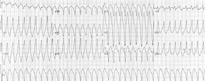
After several months and discussions with the patient and her parents, in November 1991, it was decided to insert an AICD. Five days postoperatively, a repeated electrophysiologic study was done to check the function of the device. At this time, monomorphic ventricular tachycardia was stimulated. Following appropriate sensing, the AICD delivered a 10-J shock with immediate conversion to normal sinus rhythm.
A repeat multistage exercise tolerance test was performed and was abbreviated because of patient fatigue. The patient remained in sinus rhythm throughout with appropriate sinus tachycardia to 160 beats/min during stage 3 of the modified Bruce protocol. The patient was discharged from the hospital with plans to examine her every 3 months and to obtain another biopsy specimen in 6 months. Plans were also made to follow up other family members.
DISCUSSION
Right ventricular cardiomyopathy or Fontaine's ARVD has been documented as a rare cause of sudden death.[1,2,3,8] It has been shown to have a genetic pattern that is autosomal dominant with variable penetrance and variable expression.[8]
In the literature, biopsies performed to establish the diagnosis of right ventricular dysplasia followed the demonstration of significant clinical abnormalities in the form of abnormal angiographic and echocardiographic studies and, often, documented ventricular arrhythmias.[3,9-11] The pathologic appearance of ARVD was originally described by Fontaine et al[12] and by Marcus et al.[4] These studies and subsequent series[1,3,10] have defined the fully developed lesion as being massive replacement of the right ventricular wall by fibrous and/or fatty tissue. There is a variable association with focal myocardial changes (necrosis, degeneration, or hypertrophy) and chronic inflammatory infiltrates. The process is most severe in the subepicardium and appears to move progressively toward the subendocardium. Mild focal lesions have been seen accompanying a more normal clinical picture.[10] Septal and left ventricular involvement are occasionally noted.[12,13] A pathologic entity known as fatty infiltration of the heart may confuse the diagnosis. Fatty infiltration is characterized by accumulation of lipids in the interstitium of the myocardium, particularly within the right ventricle.[14] Fatty infiltration may be distinguished from ARVD by several features, including the interstitial location of the fatty tissue, and the absence of inflammation, myofiber degeneration, or fibrosis. These differential points may not be evident on a minute endomyocardial biopsy specimen. Despite certain pathologic similarities, fatty infiltration is generally believed to lack the clinical significance associated with right ventricular dysplasia.
The presence of focal fibrosis in this biopsy specimen, although nonspecific, raises the level of suspicion of right ventricular dysplasia. In the clinical setting of the patient's family history and near-syncopal episode, the findings of the biopsy specimen, although not definitive, suggest that ARVD may be a serious possibility.
Although obtaining an endomyocardial biopsy specimen has been recommended in making the diagnosis of right ventricular dysplasia when the condition is suspected but results of clinical studies are normal,[8,10] the late involvement of the subendocardial myocardium by the process compromises the sensitivity of this procedure. This difficulty is exacerbated when evaluating the endomyocardial biopsy specimen of a young patient, where histologic evidence of what is believed to be a progressive disease can be expected to be less advanced in the subendocardium.[3]
Treatment of ARVD has been pharmacologic and/or surgical.[4-7] The use of AICD to prevent sudden death secondary to tachyarrhythmias has been demonstrated.[15,16] Implantation of this device may also be a suitable option for patients with ARVD.
Issues were raised regarding this patient and her family. First, do the benefits of placing this in our patient outweigh the disadvantages, especially when a definitive diagnosis remains elusive? Second, if all other family members have normal results of noninvasive studies, does this mean they all need endomyocardial biopsies? The answer to the first question is if the AICD functions properly, this should prevent sudden death and the risk of implantation is minimal given normal ventricular function. The second question is of a more subjective nature. It seems that the cardiomyopathy in this family manifests in the second decade of life. While all family members will be evaluated with an echocardiogram semiannually, the youngest sibling, now 12 years old, will probably be evaluated additionally with a myocardial biopsy at age 15 years.
ADDENDUM
Ten months following insertion of the AICD, the patient jogged approximately one mile, at which time she felt faint and the AICD fired three countershocks as programmed. The patient responded normally thereafter. A review of the programming of the AICD revealed no evidence of malfunction. Exercise tolerance testing could not provoke an arrhythmia.
REFERENCES
[1] Thiene G, Nava A, Corrado D, Rossi L, Pennelli N. Right ventricular cardiomyopathy and sudden death in young people. N Engl J Med 1988; 318:129-33
[2] Nava A, Scognamiglio R, Thiene G, Canciani B, Paliento L, Buja G, et al. A polymorphic form of familial arrhythmogenic right ventricular dysplasia. Am J Cardiol 1987; 59:1405-09
[3] Nava A, Thiene G, Canciani B, Scognamiglio R, Paliento L, Buja G, et al. Familial occurrence of right ventricular dysplasia: a study involving nine families. J Am Coll Cardiol 1988; 12:1222-28
[4] Marcus FI, Fontaine GH, Guiraudon GM, Frank R, Laurenceau JL, Malergue C, et al. Right ventricular dysplasia: a report of 24 adult cases. Circulation 1982; 65:384-98
[5] Fontaine G, Guiraudon G, Frank R, Tereau Y, Pavie A, Cabrol C, et al. Management of ventricular tachycardia not related to myocardial ischemia. Clin Prog Pacing Electrophysiol 1984; 2:193-219
[6] Lemery R, Brugada P, Janssen J, Cheriex E, Dugernier T, Wellens HJJ. Nonischemic sustained ventricular tachycardia: clinical outcome in 12 patients with arrhythmogenic right ventricular dysplasia. J Am Coll Cardiol 1989; 14:96-105
[7] Guiraudon GM, Klein GJ, Gulamhusein SS, Painvin GA, Del Campo C, Gonzalez JC, et al. Total disconnection of the right ventricular free wall: surgical treatment of right ventricular tachycardia associated with right ventricular dysplasia. Circulation 1983; 67:463-70
[8] Perloff JK. Familial occurrence of right ventricular dysplasia: a study involving nine families. J Am Coll Cardiol 1988; 12:1229-30
[9] Blomstrom-Lundqvist C, Selin K, Jonsson R, Johansson SR, Schlossman D, Olsson SB. Cardioangiographic findings in patients with arrhythmogenic right ventricular dysplasia. Br Heart J 1988; 59:556-63
[10] Martini B, Nava A, Thiene G, Buja GF, Canciani B, Miraglia G, et al. Accelerated idioventricular rhythm of infundibular origin in patients with concealed form of arrhythmogenic right ventricular dysplasia. Br Heart J 1988; 59:564-71
[11] Robertson JH, Bardy GH, German LL, Gallagher JJ, Kisslo J. Comparison of two dimensional echocardiographic and angiographic findings in arrhythmogenic right ventricular dysplasia. Am J Cardiol 1985; 55:1506-08
[12] Fontaine G, Guiraudon G, Frank R. Mechanism of ventricular tachycardia with and without associated chronic myocardial ischemia: surgical management based on epicardial mapping. In: Narula OS, ed. Cardiac arrhythmias: electrophysiology, diagnosis and management. Baltimore: Williams & Wilkins, 1979; 516-45
[13] Bharati S, Feld AW, Bauernfeind R, Kattus AA, Lev M. Hypoplasia of the right ventricular myocardium with ventricular tachycardia. Arch Pathol Lab Med 1983; 107:2499-2503
[14] Cotran RS, Kumar V, Robbins SL. Robbins pathologic basis of disease, 4th ed. Philadelphia: WB Saunders Company, 1989; 23
[15] Tomaselli G, Guarnieri T. The technique of automatic implantable cardioversion/defibrillation. J Crit Illness 1988; 3:87-95
[16] Mirowski M, Reid PR, Mower MM, Watkins L, Gott VL, Schauble JF, et al. Termination of malignant ventricular arrhythmias with an implanted automatic defibrillator in human beings. N Engl J Med 1980; 303:322-24
COPYRIGHT 1993 American College of Chest Physicians
COPYRIGHT 2004 Gale Group


