We report a case of bradycardia-associated torsade de pointes in which the underlying long-QT syndrome appeared to be attributable to primary cardiac conducting system disease. Our patient presented complaining of presyncope and syncope. Serial electrocardiograms obtained over a period of 10 years demonstrated slowly progressive conduction system abnormalities, and evaluation revealed no other cause. The patient's dysrhythmia was refractory to magnesium but abated with cardiac pacing at a moderate rate. A review of the relevant literature on congenital and acquired long-QT syndrome is included.
Case Report
The patient was the 74-year-old wife of a retired noncommissioned officer, who presented to the emergency department of a military treatment facility by privately owned vehicle, complaining of two episodes of dizziness and one episode of syncope. The patient's episodes of dizziness both occurred during periods of minimal exertion, while the patient was walking in her home. The first episode of dizziness occurred the day before presentation and the second occurred earlier on the day of presentation. The patient described the episodes as "lightheadedness" and a presyncopal sensation, which resolved within a few seconds of sitting down. The episode of syncope occurred the evening before presentation, while the patient was exercising on a cross-country ski simulator in her home. The episode was witnessed by her husband, who stated that it caused her to fall off the machine. It was preceded by a very brief presyncopal sensation, similar to the other two episodes she described. According to her husband, her unconsciousness lasted "a few minutes." Because the episode was brief and she recovered her baseline function quickly, appeared uninjured, and felt well thereafter, the patient did not seek medical attention at that time. Her husband stated that the patient had no convulsive activity, tongue biting, or incontinence with the syncopal episode. The patient denied chest pain, shortness of breath, or palpitations with any of the episodes. She had a history of hypertension and cigarette smoking but denied any history of heart disease, diabetes mellitus, stroke, or other major medical problems. She had no known drug allergies, and her only medications were aspirin, ramipril, and estrogen/progesterone hormone replacement. She stated that she exercised for 30 to 60 minutes several times per week with her home equipment and had never experienced symptoms with exercise before the day of presentation. During the initial evaluation, the patient said that she felt completely well and she denied symptoms of any kind.
The patient's initial vital signs were blood pressure of 166/77 mm Hg, pulse of 51 beats per minute (regular), respiratory rate of 20 breaths per minute, and temperature of 97.6°F. Room air oxygen saturation was 100%. On physical examination, the patient was alert and completely oriented. No jugular venous distension was noted. Auscultation of the lungs was remarkable for minimal bibasilar crackles. Cardiac auscultation revealed bradycardia with a rate in the low 50s and regular rhythm; no murmurs or gallops were noted. The abdomen was soft and nontender, without a palpable mass or organomegaly. The extremities were with normal peripheral pulses throughout, with no cyanosis or edema. The neurological examination indicated no deficits.
The initial electrocardiogram (ECG) (Fig. 1) showed a sinus rhythm with a ventricular rate of 44 beats per minute, first-degree atrioventricular block, right bundle branch block, and corrected QT interval (QTc) of 596 milliseconds. Compared with an ECG dated approximately 1 year previously, the morphological features and rate were essentially the same, although the QTc on the old tracing was 470 milliseconds. An ECG from ~4.5 years previously (Fig. 2) demonstrated a sinus rhythm with a rate of 53 beats per minute, PR interval of 204 milliseconds, nonspecific intraventricular conduction delay, prominent U waves, and QTc of 417 milliseconds. An ECG from 10 years previously (Fig. 3) showed the same nonspecific intraventricular conduction delay morphological features and U waves but was otherwise normal. Initial laboratory values are given in Table I. The initial chest X-ray was remarkable for slightly increased bibasilar interstitial markings but no cardiomegaly or other abnormalities.
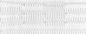
While in the emergency department, the patient was noted to have several runs of nonsustained ventricular tachycardia, which reproduced the patient's lightheadedness and presyncopal sensation but did not cause chest pain or palpitations. At first, the runs lasted for 4 or 5 beats and appeared monomorphic. The runs occurred once every 1 to 2 minutes. The patient had a palpable radial pulse during these intervals and was completely awake and conversant during the episodes, although she complained of dizziness. The patient was given 100 mg of lidocaine intravenously, and a lidocaine drip was started; the runs of ventricular tachycardia continued. The patient then had a long run of ventricular tachycardia, the rhythm strip for which is shown in Figure 4. During this episode, the undulating QRS amplitude classic for torsade de pointes (TdP) was identified. Lidocaine treatment was discontinued, and the patient was given magnesium sulfate (2 g) intravenously. Despite this, the runs of ventricular tachycardia persisted.
At this point, the patient was transferred to the intensive care unit. Central venous access was obtained via a 6-French introducer sheath in the right internal jugular vein. A balloon-guided, transvenous pacemaker was placed in the standard fashion and, when capture was noted, the patient was successfully paced from the right ventricle at a rate of 70 beats per minute, with minimal voltage, which produced the characteristic left bundle branch block QRS complexes. This completely abolished the runs of ventricular tachycardia, and no further antidysrhythmic medications were needed. A cardiac catheterization with coronary angiography was performed on hospital day 2 and revealed normal coronary arteries. A permanent pacemaker was placed on hospital day 5, and the patient was discharged home the following day, in stable condition. No symptoms were noted by the patient at the follow-up evaluation.
Discussion
Detection of long-QT syndrome (LQTS) and other entities causing sudden cardiac death (SCD) is a topic of interest to the military, given the increasing intensity of training for military readiness and also the increasing number of retirees being cared for in military hospitals around the world. Strenuous activity and conditions associated with advancing age are two instigators of fatal arrhythmias among those with LQTS.1
SCD is a common problem in the United States, with an incidence reported to be >400,000 cases per year.2 It is estimated that up to 40% of deaths occur with the first cardiac event, making this disease difficult to prevent.3 Interest in identifying the underlying cause of death has been focused on cases of unexpected arrhythmogenic death, which are estimated to represent 5% of all sudden deaths. The most prevalent causes of SCD among youths are hypertrophic obstructive cardiomyopathy, arrhythmogenic right ventricular dysplasia, Wolff-Parkinson-White syndrome, anomalous origin of the coronary arteries, and LQTS.1 In the elderly population, there is a predisposition to acquired LQTS, because diseases manifest themselves with age and patients often require potentially QT-lengthening pharmacological therapy.
In cases of SCD in which no structural heart disease can be identified, LQTS and ventricular preexcitation are the most likely causes. LQTS can be idiopathic, iatrogenic/pharmacological, or congenital. LQTS is the ECG manifestation of prolonged electrical repolarization of the cardiac muscle after the action potential. As the QT interval lengthens, the likelihood of electrical depolarization during the QT interval increases. If such an event occurs (usually attributable to a premature ventricular contraction), this may set off the reentrant polymorphic ventricular tachycardia known as TdP. TdP, if brief, may be asymptomatic, or it may present as palpitations, chest pain, lightheadedness, syncope, or SCD. An unknown population is affected by asymptomatic, periodic, prolonged QT intervals, although only a small fraction of these people go on to develop symptoms or arrhythmic complications. It is reported that 4,000 people die each year in the U.S. from TdP, with a slightly greater proportion of women being affected.4
Romano-Ward syndrome describes a prolongation of the QTc without associated noncardiac abnormalities, with an autosomal dominant genetic predilection. This is the most common form of inherited LQTS, with an estimated incidence of 1 case per 5,000 to 10,000 live births. Jervell-Lange Nielsen syndrome is a prolongation of the QTc associated with sensorineural deafness and carries a worse prognosis than Romano-Ward syndrome. The sensorineural deafness is autosomal recessive, whereas the LQTS is inherited as an autosomal dominant trait. Cases are usually found in consanguineous families, although nonconsanguineous families with Jervell-Lange Nielsen syndrome were recently identified.5
LQTS was recognized early in the 20th century, and over the past 70 years many milestones have been reached in the understanding of the underlying pathophysiological mechanisms (Table II).6 Recent advances led to recognition of the complete sixgene profile of LQTS, the five ion channels the genes encode, and their predicted associations with clinical presentations (Table III).7 The congenital form of LQTS results from mutation of certain genetic loci, altering the shape and duration of the cardiac action potential by interfering with the expression and/or functioning of these ion channels. Different metabolic states can be simulated and different medications used in the intracellular and extracellular media to determine how the channels behave in conditions that are out of the ordinary. As a result of studies like these, LQTS patients can benefit from knowing the sensitivities of their LQTS-causing mutations to different medications as well as other circumstances predisposing them to arrhythmia.8 These genetic studies have enormous implications not only for those known or suspected to carry the genes associated with the disease but also for those who are potential silent gene carriers with only mild mutations.9
There are numerous causes of acquired long QT intervals; in total, these are more common than the congenital syndromes. Some of the more common culprits include hypokalemia, hypomagnesemia, hypocalcemia, bradycardia with atrioventricular block, drug toxicity, congestive heart failure, congenital heart disease, ischemic heart disease, cerebrovascular disease, rheumatic heart disease, myocarditis, mitral valve prolapse, hypothyroidism, liquid protein diets, subarachnoid hemorrhage, and myocardial infarction.
Aside from correcting any of these predisposing conditions, treatment of LQTS is directed at regulating the heart rate, normalizing the QT length, and preventing early ventricular depolarizations. If the patient's syndrome is not associated with bradycardia, then first-line therapy is β-receptor blockade, typically with propranolol or nadolol. If β-receptor blockade fails or is contraindicated, then left cardiac sympathetic denervation is second-line therapy.6 Evidence for bradycardia or pause-induced malignant arrhythmias includes treatment with a pace-maker, with or without β-receptor blockade. If triple therapy with β-receptor blockade, left cardiac sympathetic denervation, and pacemaker treatment fails, then implantable cardioverter-defibrillators can be used as bridge therapy. Implantable cardioverter-defibrillators were shown in a limited group to be highly successful in preventing death from cardiac arrest after 3 years. However, an implantable cardioverter-defibrillator is indicated only if there has been documented ventricular tachycardia/ventricular fibrillation, an aborted cardiac arrest, or failed triple therapy.10 Rectifying QT prolongation with experimental therapy such as calcium entry blockers, pentisomide, or α-receptor blockers has been shown to be effective in some cases but only as last resort. In cases where, despite left cardiac sympathetic and β-receptor blockade, the patient remains symptomatic, right cardiac sympathetic denervation should be considered.11
A growing number of drugs are associated with QT prolongation and the concomitant risks of arrhythmia and SCD. Most of the drugs that cause LQTS are cytochrome P450 3A4 substrates (see Table IV for an abridged list of drugs associated with prolongation of the QT interval). It is important to note that, although pharmacological induction of LQTS does occur, often the patient has a history of a QT interval in the upper limits of the normal range before the initiation of drug therapy. It is thought that there is a predilection for these patients with a longer but still normal QT interval to have greater correlation with the development of a pathological rhythm.9
Screening for this disease has proved difficult. It was reported that the use of diagnostic tests to screen for cardiovascular abnormalities is ineffective and inefficient.12 The most prudent and effective methods of preparticipation screening for cardiovascular abnormalities are a history and physical examination and use of the diagnostic point system (Table V).13 When a population (athletes, active duty military, or the elderly) is identified for screening, the diagnostic point system may identify patients who are at greatest risk to die from an initial cardiac event because of a lengthened QT interval.
In a recent study, Ackerman et al.14 reported that certain stimuli, such as exercise or excitement, can trigger LQTS, which is why some cases of SCD have occurred among teens playing sports, despite normal resting ECG findings. Ackerman et al.14 administered increasing doses of epinephrine to patients with different forms of LQTS and to a control group, with continuous monitoring. Every patient with LQTS exhibited prolongation of their QT intervals when given epinephrine, whereas all other participants demonstrated shortening of their QT intervals. This study has many implications for the future of LQTS screening, especially in the field of military medicine, with the increasing number of young people subject to difficult training regimens in physically demanding environments.
Another approach, although not a preventative measure, is the further implementation of and public education in the use of automated external defibrillators (AEDs). AEDs have been shown in numerous studies to decrease the time interval from cardiac arrest to defibrillation, thus improving the survival rate for victims of ventricular fibrillation, TdP, and pulseless ventricular tachycardia. Recent Department of Defense initiatives have mandated increased participation of Federal Fire Service units in the conduct of prehospital emergency medical care at military and other federal installations. As reported by Gerhardt et al.,15 the development and deployment of an AED program for fire departments serving U.S. military installations is the first step for military applications of AEDs and their role in fatal cardiac dysrhythmias.
Although there are no currently reported screenings for this disorder among military recruits, there are efforts underway in the civilian community to discover why some are prone to QT prolongation with specific drug therapy that other people tolerate well. One approach in the management of acquired LQTS is to develop effective and efficient genetic screens. Another is to identify the molecular mechanisms responsible for drug-channel interactions that underlie these effects, with the goal of altering drug structures to minimize the risk of unwanted channel inhibition.16 Although the future of research in this field may not be clear and the practicality of testing is uncertain, it is certain that cardiac syndromes with predilection for lethal arrhythmias must be included in the differential diagnosis for a suddenly fatigued soldier or a syncopal retiree.
References
1. Drezner J: Sudden cardiac death in young athletes: causes, athlete's heart, and screening guidelines. Postgrad Med 2000; 108: 37-44, 47-50.
2. Lauer M: Focus on new technology: clinical management for survivors of sudden cardiac death. Permanente J 2001; 5: 18-32.
3. Kahn I: Clinical and therapeutic aspects of congenital and acquired long QT syndrome. Am J Med 2002; 112: 58-66.
4. Leon A: New guideline published-cardiac dysrhythmias-rapid rhythms. EBM Brief 2003; 3. Available at http://www.imakenews.com.ebmsolutions/e_article000156116.cfm; accessed January 15, 2004.
5. Hoorntje T, Alders M, Van Tintelen P, et al: Homozygous premature truncation of the HERO protein: the human HERG knockout. Circulation 1999; 100: 1264-7.
6. Schwartz PJ: Long QT syndrome, European information Center. Available at www.qtsyndrome.ch/timeline; accessed January 5, 2004.
7. Kubota T, Horie M, Takano M, et al: Evidence for a single nucleotide polymorphism in the KCNQ1 potassium channel that underlies susceptibility to lifethreatening arrhythmias. J Cardiovasc Eleetrophysiol 2001; 12: 1230-1.
8. Chapman H, Paavonen K, Pasternack M: Long QT syndrome background. Helsinki, Finland, Molecular Neurophysiology, Institute of Biotechnology, University of Helsinki. Available at http://www.biocenter.helsinki.fi/bi/physiol/Long%20QT/Long%20QT.htm; accessed January 6, 2004.
9. Viskin S: Long QT syndromes and torsade de pointes. Lancet 1999; 354: 1625-33.
10. Moss A: The long QT syndrome: overview and management, February 2002. Available at http://omaha.physiol.arizona.edu/Physiology485/Gruener/LongQTSyndrome.pdf; accessed January 4, 2004.
11. Zareba W, Rosero S: Long QT syndrome, updated May 10, 2002. Available at http://www.emedicine.com/med/topic1983.htm; accessed January 1, 2004
12. Koester M: A review of sudden cardiac death in young athletes and strategies for preparticipation cardiovascular screening. J Athl Train 2001; 36: 197-204.
13. Subramanian R, Venugopalan P: Congenital long QT syndromes in children. Indian Heart J 2002; 54: 152-8.
14. Ackerman M, Khositseth A, Nemec J, Hejlik J, Shen W: Effect of phenylephrine provocation on dispersion of repolarization in congenital long QT syndrome. Ann Noninvasive Electrocardiol 2003; 8: 208-14.
15. Gerhardt R, Wiegert R, Newell S, Gerhardt D, Gaither P: Field deployment of automated external defibrillators to a Federal Fire Service unit under medical direction of a military emergency service. Milit Med 1999; 164: 877-80.
16. Larsen L, Andersen P, Ranters J, et al: Screening for mutations and polymorphisms in the genes KCNH2 and KCNE2 encoding the cardiac HERG/MiRP1 ion channel: implications for acquired and congenital long Q-T syndrome. Clin Chem 2001; 47: 1390-5.
Guarantor; CPT Simeon W. Ashworth, MC USA
Contributors: CPT Simeon W. Ashworth, MC USA; CPT Marc E. Levsky, MC USA; CPT Chad T. Marley, MC USA; MAJ Christopher S. Kang, MC USA
Madigan Army Medical Center/University of Washington Emergency Medicine Residency Program, Department of Emergency Medicine, Madigan Army Medical Center, Tacoma, WA 98433.
The opinions and assertions contained herein are the private views of the authors and should not be construed as official or as reflecting the views of the Department of the Army or the Department of Defense.
This manuscript was received for review in February 2004 and was accepted for publication in June 2004.
Copyright Association of Military Surgeons of the United States May 2005
Provided by ProQuest Information and Learning Company. All rights Reserved


