* This is a case report of ascariasis of the common bile duct in a 65-year-old man from Colombia who had undergone prior cholecystectomy. The patient presented with postprandial epigastric pain and a 20-Ib weight loss. The laboratory findings were remarkable for peripheral blood eosinophilia. The ultrasound finding was suggestive of periampullary or pancreatic neoplasm. He underwent endoscopic retrograde cholangiopancreatography with endoscopic extraction of a motile, live worm identified as Ascaris lumbricoides. Roundworm infestation should always be suspected in immigrants from endemic areas who present with hepatobiliary symptoms.
(Arch Pathol Lab Med. 2000;124:1231-1232)
The first report of an adult Ascaris lumbricoides roundworm in the biliary ducts presenting clinically (and radiologically) in North America or the United Kingdom was published in 1977.1 Ascariasis is a helminthic infection of global distribution with more than 1.4 billion persons infected throughout the world. Most infections occur in the developing countries of Asia and Latin America. In the United States, ascariasis is the third most common helminth infection (exceeded only by hookworm and whipworm infections). Of 4 million people infected in the United States, a large percentage are immigrants from developing countries, with infection rates of 20% to 60%.2
REPORT OF A CASE
A 67-year-old man from Colombia presented with a 3-month history of postprandial epigastric pain and a 20-lb weight loss. He had no fever, chills, jaundice, or diarrhea. Results of his physical examination were unremarkable, with no abdominal tenderness. Laboratory findings included the following values: white blood cells, 13.6 x 10^sup 9^/L with 10% eosinophils; bilirubin, 10.3 wmol/L; gamma-glutamyltransferase, 351 U/L; alkaline phosphatase, 142 U/L; aspartate aminotransferase, 35 U/L; and alanine aminotransferase, 17 U/L. Ultrasound revealed a dilated common bile duct (1.2 cm), with abrupt termination at the distal end suggestive of a periampullary or pancreatic neoplasm. Ductal dilation was not confirmed by a computed tomographic scan, but a possible hypodense lesion was noted in the pancreatic head at the level of the papilla of Vater.
The patient underwent an endoscopic retrograde cholangiopancreatography (ERCP). Contrast injection of the common bile duct demonstrated a vermiform filling defect that extended from the distal duct into a right hepatic duct (Figure 1 ). The ducts did not appear significantly dilated. An 11.5-mm balloon catheter was introduced through the papilla and inflated alongside the filling defect. By pulling the inflated balloon down the duct, a tan worm emerged through the papilla. The balloon was deflated and removed, and the protruding worm was captured by a snare. The tan worm was retrieved intact and proved to be a motile live Ascaris. The duodenoscope and snare were carefully withdrawn together. The duodenoscope was reintroduced, and subsequent cholangiography was normal. The specimen was sent to the laboratory for analysis and was identified as an adult female A lumbricoides (Figure 2). A common bile duct brushing was also performed, which revealed numerous bacterial colonies and scattered Ascaris eggs. The patient was treated with mebendazole, with complete resolution of his symptoms and subsequent return to normal weight.
COMMENT
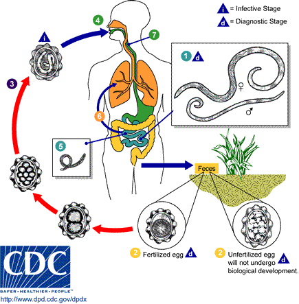
Ascariasis is a helminthic infection of humans caused by the nematode A lumbricoides. Ascaris eggs are passed in the feces. The fertilized eggs require 10 to 15 days in the soil for embryonation before they are infective. Infection follows ingestion of embryonated eggs. After ingestion, the shell of the embryonated egg is dissolved by the gastric juice, and the embryo emerges in the duodenum as the rhabdoid larva 200 to 300 (mu)m long and 13 to 15 wm thick. The larvae migrate to the cecum and penetrate the surface epithelium of the mucosa. The larvae enter the veins of the portal system and are carried to the liver. In the liver, larvae move freely in the sinusoids. Some may subsequently pass via the hepatic veins to the heart and lungs. Some larvae may enter the lymphatic system of bowel and are carried via the thoracic duct to the lungs. The larvae break through the capillary wall and enter the alveolar space and the bronchial tree. They ascend the tracheobronchial tree and larynx to the hypopharynx, where they are swallowed. On reaching the small intestine, the larvae attain sexual maturity in 2 to 3 months. The normal habitat for the adult worm is the jejunum. The time from the larval infection to maturity of adult worms is usually 4 months.2
Most Ascaris infections are without clinical disease. Clinical disease is mostly restricted to subjects with heavy worm loads. The manifestations of ascariasis vary and include constitutional symptoms, particularly pulmonary and gastrointestinal complaints.3 Hepatobiliary and pancreatic ascariasis can cause 5 distinct clinical presentations: biliary colic, acalculous cholecystitis, acute cholangitis, acute pancreatitis, and hepatic abscess.2
Sandouk et al reported that in endemic countries ascariasis should be suspected in patients with pancreatic-biliary disease, especially if a cholecystectomy or sphincterotomy has been performed in the past.4 The diagnosis is made in most cases by sampling stool for ova and parasites. Radiographic and endoscopic methods are helpful.3 In addition, ERCP can help diagnose biliary and pancreatic ascariasis, including Ascaris in the duodenum. Also, ERCP can be used to extract worms from the biliary and pancreatic ducts.2 Notably, ERCP carries less morbidity and mortality than the surgical approach.s Several drugs are effective in treatment of ascariasis. Pyrantel pamoate (a single dose of 11 mg/kg orally, with a maximum dose of 1.0 g), mebendazole (100 mg orally 2 times per day for 3 days), and albendazole (a single dose of 400 mg orally) are drugs of first choice.2 Treatment with an antihelminthic agent is usually effective in mild cases, and prognosis is excellent. Endoscopy is successful in the treatment of Ascaris infestation resistant to medical therapy.6 More severe infection may cause significant morbidity and require surgical intervention.2 Surgery is important in the management of infestations complicated by biliary or pancreatic strictures and stones or worms in the gallbladder.6
References
1. Schulman A. Biliary ascariasis presenting in the United States. Am / Gastroenterol. 1977;2:167-170.
2. Khuroo MS. Ascariasis. Gastroenterol Clin North Am. 1996;3:553-577.
3. Bratton R, Nesse R, Ascariasis. Postgrad Med. 1993;93:171-173,177-178.
4. Sandouk F, Haffar S, Zada M, Graham D, Anand B. Pancreatic-biliary ascariasis: experience of 300 cases. Am / Gastroenterol. 1997;12:2264-2267.
S. Osman M, Lausten SB, EI-Sefi T, Boghdadi I, Rashed MY, Jensen SL. Biliary parasites. Diagn Surg. 1998;4:287-296.
6. Beckingham IJ, Cullis SN, Krige JE, Bornman PC, Terblanche ). Management of hepatobiliary and pancreatic Ascaris infestation in adults after failed medical treatment. Br J Surg. 1998;7:907-910.
Accepted for publication December 28, 1999.
From the Departments of Pathology and Laboratory Medicine (Drs Amog, Sieber, and EI-Fanek) and Medicine (Dr Lichtenstein), Danbury Hospital, Danbury, Conn.
Reprints: Glenda Amog, MD, Department of Pathology and Laboratory Medicine, Danbury Hospital, 24 Hospital Ave, Danbury, CT 06810 (e-mail: GFAmog@aol.com).
Copyright College of American Pathologists Aug 2000
Provided by ProQuest Information and Learning Company. All rights Reserved


