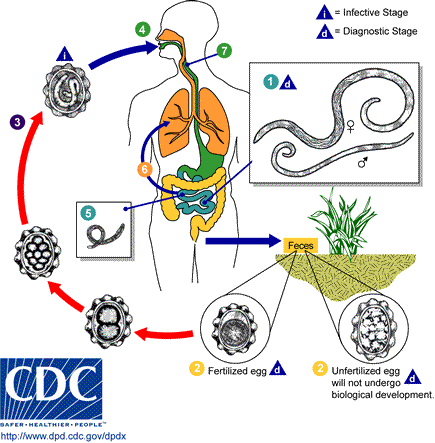
An adult female Ascaris worm.Adult worms (1) live in the lumen of the small intestine. A female may produce approximately 200,000 eggs per day, which are passed with the feces (2). Unfertilized eggs may be ingested but are not infective. Fertile eggs embryonate and become infective after 18 days to several weeks (3), depending on the environmental conditions (optimum: moist, warm, shaded soil). After infective eggs are swallowed (4), the larvae hatch (5), invade the intestinal mucosa, and are carried via the portal, then systemic circulation to the lungs . The larvae mature further in the lungs (6) (10 to 14 days), penetrate the alveolar walls, ascend the bronchial tree to the throat, and are swallowed (7). Upon reaching the small intestine, they develop into adult worms (8). Between 2 and 3 months are required from ingestion of the infective eggs to oviposition by the adult female. Adult worms can live 1 to 2 years.
Find information on thousands of medical conditions and prescription drugs.

Ascariasis

Ascariasis is a debilitating human disease caused by the roundworm Ascaris lumbricoides; other species of Ascaris are parasitic in domestic animals (see Nematode). Perhaps as many as one quarter of the world's people are infected, but ascariasis is particularly prevalent in tropical regions and in areas of poor hygiene. more...

Home
Diseases
A
Aagenaes syndrome
Aarskog Ose Pande syndrome
Aarskog syndrome
Aase Smith syndrome
Aase syndrome
ABCD syndrome
Abdallat Davis Farrage...
Abdominal aortic aneurysm
Abdominal cystic...
Abdominal defects
Ablutophobia
Absence of Gluteal muscle
Acalvaria
Acanthocheilonemiasis
Acanthocytosis
Acarophobia
Acatalasemia
Accessory pancreas
Achalasia
Achard syndrome
Achard-Thiers syndrome
Acheiropodia
Achondrogenesis
Achondrogenesis type 1A
Achondrogenesis type 1B
Achondroplasia
Achondroplastic dwarfism
Achromatopsia
Acid maltase deficiency
Ackerman syndrome
Acne
Acne rosacea
Acoustic neuroma
Acquired ichthyosis
Acquired syphilis
Acrofacial dysostosis,...
Acromegaly
Acrophobia
Acrospiroma
Actinomycosis
Activated protein C...
Acute febrile...
Acute intermittent porphyria
Acute lymphoblastic leukemia
Acute lymphocytic leukemia
Acute mountain sickness
Acute myelocytic leukemia
Acute myelogenous leukemia
Acute necrotizing...
Acute promyelocytic leukemia
Acute renal failure
Acute respiratory...
Acute tubular necrosis
Adams Nance syndrome
Adams-Oliver syndrome
Addison's disease
Adducted thumb syndrome...
Adenoid cystic carcinoma
Adenoma
Adenomyosis
Adenosine deaminase...
Adenosine monophosphate...
Adie syndrome
Adrenal incidentaloma
Adrenal insufficiency
Adrenocortical carcinoma
Adrenogenital syndrome
Adrenoleukodystrophy
Aerophobia
Agoraphobia
Agrizoophobia
Agyrophobia
Aicardi syndrome
Aichmophobia
AIDS
AIDS Dementia Complex
Ainhum
Albinism
Albright's hereditary...
Albuminurophobia
Alcaptonuria
Alcohol fetopathy
Alcoholic hepatitis
Alcoholic liver cirrhosis
Alektorophobia
Alexander disease
Alien hand syndrome
Alkaptonuria
Alliumphobia
Alopecia
Alopecia areata
Alopecia totalis
Alopecia universalis
Alpers disease
Alpha 1-antitrypsin...
Alpha-mannosidosis
Alport syndrome
Alternating hemiplegia
Alzheimer's disease
Amaurosis
Amblyopia
Ambras syndrome
Amelogenesis imperfecta
Amenorrhea
American trypanosomiasis
Amoebiasis
Amyloidosis
Amyotrophic lateral...
Anaphylaxis
Androgen insensitivity...
Anemia
Anemia, Diamond-Blackfan
Anemia, Pernicious
Anemia, Sideroblastic
Anemophobia
Anencephaly
Aneurysm
Aneurysm
Aneurysm of sinus of...
Angelman syndrome
Anguillulosis
Aniridia
Anisakiasis
Ankylosing spondylitis
Ankylostomiasis
Annular pancreas
Anorchidism
Anorexia nervosa
Anosmia
Anotia
Anthophobia
Anthrax disease
Antiphospholipid syndrome
Antisocial personality...
Antithrombin deficiency,...
Anton's syndrome
Aortic aneurysm
Aortic coarctation
Aortic dissection
Aortic valve stenosis
Apert syndrome
Aphthous stomatitis
Apiphobia
Aplastic anemia
Appendicitis
Apraxia
Arachnoiditis
Argininosuccinate...
Argininosuccinic aciduria
Argyria
Arnold-Chiari malformation
Arrhythmogenic right...
Arteriovenous malformation
Arteritis
Arthritis
Arthritis, Juvenile
Arthrogryposis
Arthrogryposis multiplex...
Asbestosis
Ascariasis
Aseptic meningitis
Asherman's syndrome
Aspartylglycosaminuria
Aspergillosis
Asphyxia neonatorum
Asthenia
Asthenia
Asthenophobia
Asthma
Astrocytoma
Ataxia telangiectasia
Atelectasis
Atelosteogenesis, type II
Atherosclerosis
Athetosis
Atopic Dermatitis
Atrial septal defect
Atrioventricular septal...
Atrophy
Attention Deficit...
Autoimmune hepatitis
Autoimmune...
Automysophobia
Autonomic dysfunction
Familial Alzheimer disease
Senescence
B
C
D
E
F
G
H
I
J
K
L
M
N
O
P
Q
R
S
T
U
V
W
X
Y
Z
Medicines

Infection occurs through ingestion of food contaminated with fecal matter containing Ascaris eggs. The larvae hatch, burrow through the intestine, reach the lungs, and finally migrate up the respiratory tract. From there they are then reswallowed and mature in the intestine, growing up to 30 cm (12 in.) in length and anchoring themselves to the intestinal wall.

Infections are usually accompanied by inflammation, fever, and diarrhea, and serious problems may develop if the worms migrate to other parts of the body.

Prevalence

Roughly 1.5 billion individuals are infected with this worm1. Ascariasis is endemic in the United States including Gulf Coast and Ozark Mountains; in Nigeria and in Southeast Asia. One study indicated that the prevalence of ascariasis in the United States at about 4 million (2%). In a survey of a rural Nova Scotia community, 28.1% of 431 individuals tested were positive for Ascaris, all of them being under age 20, while all 276 tested in metropolitan Halifax were negative2.

Deposition of ova (eggs) in sewage hints at the degree of ascariasis incidence. A 1978 study showed about 75% of all sewage sludge samples sampled in United States urban catchments contained Ascaris ova, with rates as high as 5 to 100 eggs per liter. In Frankfort, Indiana, 87.5% of the sludge samples were positive with Ascaris, Toxocara, Trichuris, and hookworm. In Macon, Georgia, one of the 13 soil samples tested positive for Ascaris. Municipal wastewater in Riyadh, Saudi Arabia detected over 100 eggs per liter of wastewater 3 and in Czechoslovakia was as high as 240-1050 eggs per liter 4.

Ascariasis sources can often be measured by examining food for ova. In one field study in Marrakech, Morocco, where raw sewage is used to fertilize crop fields, Ascaris eggs were detected at the rate of 0.18 eggs/kg in potatoes, 0.27 eggs/kg in turnip, 4.63 eggs/kg in mint, 0.7 eggs/kg in carrots, and 1.64 eggs/kg in radish5. A similar study in the same area showed that 73% of children working on these farms were infected with helminths, particularly Ascaris, probably as a result of exposure to the raw sewage.

Life cycle

First appearance of eggs in stools is 60-70 days. In larval ascariasis, symptoms occur 4-16 days after infection. The final symptoms are gastrointestinal discomfort, colic and vomiting, fever; observation of live worms in stools. Some patients may have pulmonary symptoms or neurological disorders during migration of the larvae. However there are generally few or no symptoms. A bolus of worms may obstruct the intestine; migrating larvae may cause pneumonitis and eosinophilia.

Read more at Wikipedia.org


[List your site here Free!]


Intestinal Spirochetosis
From Archives of Pathology & Laboratory Medicine, 7/1/04 by Nodit, Laurentia

The patient was a 9-year-old boy with recurrent abdominal pain sometimes associated with diarrhea and hematochezia. Extensive laboratory investigation was unremarkable and colonoscopy showed only mild erythema of the rectal mucosa.

The histologic evaluation of the colonic mucosa revealed a 2- to 3-µm-thick band of spirochetes that covered the luminal surface of the absorptive cells (Figure 1) and spared goblet cells (Figure 2). There was no significant inflammation in the lamina propria except for a mild increase in the number of eosinophils in places (Figure 1). The upper gastrointestinal tract showed no pathologic changes.

The patient was treated with erythromycin, 40 mg/kg/ day, for 10 days. With treatment, his abdominal pain resolved and rectal bleeding ceased.

Intestinal spirochetosis is characterized by heavy colonization of colonic mucosa by spirochetes. The presence of spirochetes in human bowel has been recognized since 1900, but it is still a matter of debate whether they are just commensal organisms or whether they represent a true colorectal disease.1

Human intestinal spirochetes comprise a group of bacteria of considerable heterogeneity, with many of them being classified as Borrelia eurygyrata, Brachyspirn aalborgi, or Serpulina pilosicoli. They are genetically unrelated to non-intestinal spirochetes, such as Treponema pallidum. A heavy infestation by intestinal spirochetes is described in both immunocompetent and immunocompromised hosts.

The mode of transmission remains unknown. Chronic fecal stasis might be implicated in spirochetal multiplication. In children, intestinal spirochetosis has been associated with ascariasis, enterobiasis, and Helicobacter pylori gastritis. Whether this is coincidental or reflects an oral route of infection is unclear.

Many authors regard these organisms as commensals, because small numbers of spirochetes are usually not associated with clinical symptoms. A heavy colonization, however, histologically appearing as a dense basophilic band along the entire colonic surface, may be associated with diarrhea, rectal bleeding, abdominal pain, purulent discharge, and an appendicitis-like picture. Symptoms can persist even after the eradication of spirochetes.

The spirochetes appear on hematoxylin-eosin-stained sections as a densely packed layer of organisms, which covers the luminal surface of the colonic absorptive epithelial cells but spares the goblet cells. The presence of intestinal spirochetes has been also documented within the crypt epithelial cells and lamina propria histiocytes as well as within Schwann cells. These findings reflect a potential for invasion and cell damage. The colonization is usually associated with no or only minimal inflammatory reaction within the lamina propria. The intestinal spirochetes are highlighted by periodic acid-Schiff, Giemsa, and Grocott as well as silver stains such as Warthin-Starry (Figure 3) and Dieterle.

A number of diagnostic tests based on genotypes have been developed for identification of Brachyspira species, including specific detection by polymerase chain reaction targeting 16S ribosomal DNA or 23S rDNA and fluorescent rRNA in situ hybridization with specific oligonucleotide probes directly from feces or paraffin-embedded tissue. There is no clear association between species and clinical or pathologic findings.2

References

1. Rotterdam H. Intestinal spirochetosis. In: Connor DH, Chandler FW, Schwartz DA, Manz HJ, Lack EE, eds. Pathology of Infectious Diseases. Stamford, Conn: Appleton & Lange; 1997:583-588.

2. Koteish A, Kannangai R, Abraham S, Torbenson M. Colonic spirochetosis in children and adults. Am J Clin Pathol. 2003;120:828-832.

Laurentia Nodit, MD; Maria Parizhskaya, MD

Accepted for publication March 10, 2004.

From the Department of Pathology, University of Pittsburgh Medical Center and Children's Hospital, Pittsburgh, Pa.

The authors have no relevant financial interest in the products or companies described in this article.

Reprints: Laurentia Nodit, MD, Department of Pathology, University of Pittsburgh Medical Center, A-610 Scaife Hall, 200 Lothrop St, Pittsburgh, PA 15213 (e-mail: noditl@upmc.edu).

Copyright College of American Pathologists Jul 2004
Provided by ProQuest Information and Learning Company. All rights Reserved

Return to Ascariasis
Home Contact Resources Exchange Links ebay