Definition
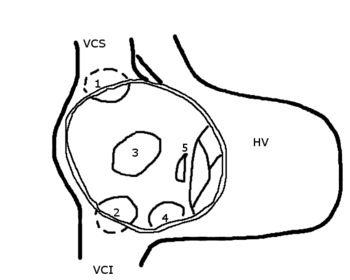
An atrial septal defect is an abnormal opening in the wall separating the left and right upper chambers (atria) of the heart.
Description
During the normal development of the fetal heart, there is an opening in the wall (the septum) separating the left and right upper chambers of the heart. Normally, this opening closes before birth, but if it does not, the child is born with a hole between the left and right atria. This abnormal opening is called an atrial septal defect and causes blood from the left atrium to flow into the right atrium.
Different types of atrial septal defects can occur, and they are classified according to where in the separating wall they are found. The most commonly found atrial septal defect occurs in the middle of the atrial septum and accounts for about 70% of all atrial septal defects. Abnormal openings can form in the upper and lower parts of the atrial septum as well.
Causes & symptoms
Abnormal openings in the atrial septum occur during fetal development and are twice as common in females as in males. These abnormalities can go unnoticed if the opening is small, producing no abnormal symptoms. If the defect is big, large amounts of blood flowing from the left to the right atrium will cause the right atrium to swell to hold the extra blood.
People born with an atrial septal defect can have no symptoms through their twenties, but by age 40, most people with this condition have symptoms that can include shortness of breath, rapid abnormal beating of the atria (atrial fibrillation), and eventually heart failure.
Diagnosis
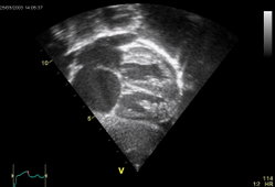
Atrial septal defects can be identified by various methods. Abnormal changes in the sound of the heart beats can be heard when a doctor listens to the heart with a stethoscope. In addition, a chest x ray, an electrocardiogram (ECG, an electrical printout of the heartbeats), and an echocardiogram (a test that uses sound waves to form a detailed image of the heart) can also be used to identify this condition.
An atrial septal defect can also be diagnosed by using a test called cardiac catheterization. This test involves inserting a very thin tube (catheter) into the heart's chambers to measure the amount of oxygen present in the blood within the heart. If the heart has an opening between the atria, oxygen-rich blood from the left atrium enters the right atrium. Through cardiac catheterization, doctors can detect the higher-than-normal amount of oxygen in the heart's right atrium, right ventricle, and the large blood vessels that carry blood to the lungs, where the blood would normally subsequently get its oxygen.
Treatment
Atrial septal defects often correct themselves without medical treatments by the age of two. If this dose not happen, surgery is done by sewing the hole closed, or by sewing a patch of Dacron material or a piece of the sac that surrounds the heart (the pericardium), over the opening.
Some patients can have the defect fixed by having an clam-shaped plug placed over the opening. This plug is a man-made device that is put in place through a catheter inserted into the heart.
Prognosis
Individuals with small defects can live a normal life, but larger defects require surgical correction. Less than 1% of people younger than 45 years of age die from corrective surgery. Five to ten percent of patients can die from the surgery if they are older than 40 and have other heart-related problems. When an atrial septal defect is corrected within the first 20 years of life, there is an excellent chance for the individual to live normally.
Key Terms
- Cardiac catheterization
- A test that involves having a tiny tube inserted into the heart through a blood vessel.
- Dacron
- A synthetic polyester fiber used to surgically repair damaged sections of heart muscle and blood vessel walls.
- Echocardiogram
- A test that uses sound waves to generate an image of the heart, its valves, and chambers.
Further Reading
For Your Information
Books
- "Congenital Heart Disease." In Texas Heart Institute Heart Owner's Handbook. New York: John Wiley & Sons, Inc., 1996, pp.259-263.
- "If Your Child Has A Congenital Heart Defect." In Cardiovascular Diseases and Disorders Sourcebook, edited by Karen Bellenir and Peter D. Dresser. Detroit: Omnigraphics, Inc., 1995, p.216.
- Friedman, William F., and John S. Child. "Congenital Heart Disease in the Adult." In Harrison's Principles of Internal Medicine, edited by Anthony S. Fauci, et al. New York: McGraw Hill, 1998, pp.1303-1304.
- Nugent, Elizabeth W., et al. "The Pathology, Pathophysiology, Recognition, and Treatment of Congenital Heart Disease." In Hurst's The Heart, edited by Robert C. Schlant, et al. New York: McGraw Hill, 1994, pp. 1773-1777.
Organizations
- The American Heart Association. 7272 Greenville Ave., Dallas, TX 75231-4596. (800)787-8984. http://www.americanheart.org/.
Gale Encyclopedia of Medicine. Gale Research, 1999.


