To the Editor:
Raised concentrations of cardiac troponin, indicating poor short-term prognosis, have been reported in patients with dilated and secondary cardiomyopathy. (1) It is assumed that these elevated levels of troponin reflect ongoing myocyte degeneration. Heavy-resistance exercise and strength training are associated with adaptive changes of cardiac structure and function. (2) Furthermore, recently published data demonstrate that more than one third of patients who received a clinical diagnosis of pulmonary embolism had presented with elevated serum troponin concentrations due to minor myocardial damage in acute right ventricular dysfunction. (3)
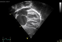
We report on a 71-year-old man with a 9-year history of ostium secundum type-atrial septal defect and increased right ventricular size. The patient was admitted to our hospital because of dyspnea and chest discomfort after heavy-resistance exercise, moving furniture. The patient's BP was 120/80 mm Hg, his pulse rate was 82 beats/min, and his breathing rate was 18 breaths/min. The first heart sound was loud, the second heart sound was widely split without widening during inspiration, and a pulmonary systolic ejection murmur was present. ECG revealed complete right bundle-branch block and signs of right ventricular hypertrophy. No changes in the repolarization pattern could be documented compared with ECGs recorded previously. Echocardiography showed increased right ventricular size, moderate right ventricular dysfunction, and tricuspid regurgitation, indicative of increased right ventricular stroke volume. A pulmonary-to-systemic flow ratio of 2:1, due to an ostium secundum type-atrial septal defect, could be accurately diagnosed by Doppler echocardiography. Symptoms disappeared spontanously within a few minutes after hospital admission without therapy.
Laboratory examination revealed elevated serum concentrations of cardiac troponin I (2.2 ng/mL; normal < 1.6 ng mL) and myocardial band (MB) isoenzyme of creatine kinase (13 U/L; normal < 5 U/L). MB isoenzyme of creatine kinase peaked (17 U/L) 4 h after hospital admission, and the level of troponin I raised to a peak concentration of 6.4 ng/mL. Other laboratory findings, including inflammatory markers and coagulation tests, were within normal ranges. Coronary angiography showed normal epicardial coronary vessels with no signs of atherosclerotic lesions, and a cineangiogram revealed no wall motion abnormalities.
For several reasons, we do not believe that ischemia caused the elevation of cardiac enzymes. First, a normal coronary angiogram finding does not definitely rule out previous coronary artery occlusion, but it makes myocardial ischemia or myocardial infarction unlikely, particularly in the absence of known risk factors for atherosclerosis. (4,5) Second, neither echocardiography nor cineangiography revealed any wall-motion abnormalities indicative of segmental myocardial damage, Third, elevated concentrations of cardiac troponin have been reported in patients with dilated cardiomyopathy, secondary cardiomyopathies, and acute right ventricular dysfunction. (1,3)
To our knowledge, this is the first report in which heavy-resistance exercise induced a raise of levels of serum cardiac troponin and MB isoenzyme of creatine kinase in a patient with preexisting dilatation and dysfunction of the right ventricle due to a ostium secundum type-atrial septal defect with left-to-right shunt.
The management of atrial septal defects in older patients is debated persistently, and clinical trials (6,7) have demonstrated conflicting data. We do not have absolute proof to guide our decisions; therefore, we must rely on our experience as well as on the available evidence in deciding how to treat these patients. (8) Further studies are needed to determine whether the management of these patients should be guided by clinical signs or by biochemical markers, like troponins.
Correspondence to: Johann Auer, MD, Second Medical Department, Division of Cardiology and Intensive Care; General Hospital Wels, Grieskirchnerstra[beta]e 42, A-4600 Wels, Austria; e-mail: johann.auer@khwels.at
REFERENCES
(1) Sato Y, Yamada T, Taniguchi R, et al. Serum concentration of cardiac troponin T inpatients with cardiomypoathy: a possible mechanism of acute heart failure. Heart 1998; 80:209-210
(2) Pluim BM, Zwinderman AH, van der Laarse A, et al. The athlete's heart: a meta-analysis of cardiac structure and function. Circulation 1999; 100:336-344
(3) Meyer T, Binder L, Hruska N, et al. Cardiac troponin I elevation in acute pulmonary embolism is associated with right ventricular dysfunction. J Am Coll Cardiol 2000; 36:1632-1636
(4) Cheitlin MD, Sokolow M, McIlroy MB. Coronary heart disease. In: Cheitlin MD, Sokolow M, McIlroy MB, eds. Clinical cardiology, 6th ed. Norwalk, CT: Appleton and Lange, 1993; 181
(5) Michaelson SP, Karsh DL, Wolfson S, et al. Recurrent myocardial infarction with normal coronary arteriography. N Engl J Med 1977; 297:916-917
(6) Shah D, Azhar M, Oakley CM, et al. Natural history of secundum atrial septal defect in adults after medical or surgical treatment: a historical prospective study. Br Heart J 1994; 71:224-227
(7) Jemielity M, Dyszkiewicz L, Perek B, et al. Do patients over 40 years of age benefit from surgical closure of atrial septal defects? Heart 2001; 85:300-303
(8) Webb G. Do atients over 40 ears of a e benefit from closure of an atrial septal defect. Heart 2001; 85:249-250
COPYRIGHT 2001 American College of Chest Physicians
COPYRIGHT 2001 Gale Group


