The thin-walled right ventricle (RV) has received little attention in the past compared with its muscular neighbor, the left ventricle (LV).[1,2] Recently, the availability of better bedside techniques to study RV function, such as 2D echocardiography, radionuclear angiography, and the fast-response thermistor catheter, have allowed us to understand the role of the RV in many clinical settings. RV dysfunction has been implicated in a spectrum of cardiopulmonary disorders including pulmonary embolism, COPD, ARDS, application of PEEP, sepsis, myocardial contusion, and response to cardiopulmonary bypass.[2] Physiologic abnormalities of the RV that may lead to dysfunction or failure include pressure overload, volume overload, and diminished contractility. This editorial will focus on RV dysfunction associated with pressure overload.
In acute pulmonary hypertension, irrespective of the etiology, RV stroke work (RVSW) increases to counter the elevated pulmonary vascular resistance (PVR). With progressive increases in PVR, RVSW begins to decline. This fall in RVSW is generally seen in a fall of RV stroke volume and diminished left ventricular preload.[3]
The principles for right ventricular resuscitation have been proposed by Laver[4] and numerous others. Chronologically, the steps of RV resuscitation are: (1) provision of adequate RV filling volume as indicated by central venous pressure; (2) maintenance of adequate coronary perfusion pressure, particularly in the presence of RV disease or right coronary artery atherosclerosis; (3) use of vasodilators to control the capacitance bed; and (4) application of inotropic support for direct impact on ventricular contractility. Several comments are in order regarding the above guidelines for RV resuscitation. Right atrial pressure is usually increased in conditions of acute or chronic pressure overload. This leads to reduction in available venous return (VR) gradient. The VR to the right atrium (RA) is maintained by the pressure gradient between mean systemic pressure (Pms) and right atrial pressure (RAP). The Pms is the average pressure in the circulation under conditions of no flow.[5,6] Volume resuscitation increases Pms and thus available VR gradient (Pms-RAP) to the RV. The increase in Pms causes considerable elevation in peripheral venous pressure, leading to the development of peripheral edema. Under these conditions, peripheral edema is a physiologic necessity, as diuretic therapy may lead to reduction in VR and cardiac output.[6]
Overenthusiastic volume resuscitation to improve RV performance has several limitations. Volume expansion resulting in increased RV filling pressure will lower the blood flow to the RV free wall. This occurs in the face of increased RV myocardial work and oxygen demand.[7] In addition, overdistention of the RV can cause concomitant changes in geometry and compliance of the LV. This may lead to errors in interpretation and management of hemodynamic data derived from invasive monitoring.[8]
Recent experimental studies[9,10] suggest that use of vasoconstrictors may be beneficial in RV dysfunction by maintaining mean aortic pressure (MAP), which is crucial for coronary perfusion. RV myocardial perfusion is dependent on the gradient from MAP to right ventricular end-diastolic pressure (RVEDP).[11] The second critical component of right coronary perfusion is anatomic status. In some cases, systemic hypertension may be required in the presence of high pulmonary artery pressure to improve RV performance. Laver proposes use of nitroglycerin (NTG) and epinephrine or dopamine in combination to provide control of capacitance vessel tone along with reduction in elevated RV filling pressures. NTG may also recruit coronary vasodilator reserve. The effect of combined use of NTG plus a pressor on RV myocardial blood flow and RV coronary vasodilator reserve is not known at this time.
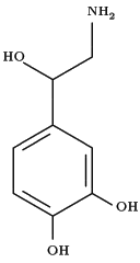
In this issue of Chest (see page 1333), Angle and coworkers, in a canine model of pulmonary embolism of sufficient severity to decrease measured cardiac output, demonstrated that norepinephrine (NE) titrated to increase the systemic blood pressure produced significant improvement in ventricular performance without compromise in renal blood flow or function. In addition, NE therapy did not increase pulmonary vascular resistance. In their discussion, the authors propose increased RV contractility and augmented right coronary perfusion as possible means for the improvement in RV performance seen. No objective data are provided, however, to permit distinction between these two potential modes of RV performance improvement.
There is considerable reluctance among intensive care physicians to use NE in the supportive management of RV dysfunction. This reluctance is based on the reported contributions to renal insufficiency associated with NE therapy. In a recent clinical study, Desjars et al[12] failed to show deterioration in renal function and described improved renal performance with the use of NE. A possible explanation is found in work demonstrating decreased proximal tubular reabsorption with increased renal perfusion pressure. In addition, there is increased sodium and water delivery to the distal tubule.[13] Schaer et al[14] in a recent experimental study observed that impairment of renal blood flow caused by NE could be reversed by simultaneous infusion of low-dose dopamine. The clinical role for NE and dopamine administration in support of RV performance and renal function remains to be established.
To summarize, in low cardiac output states complicating acute pulmonary hypertension, RV performance can be improved by careful volume resuscitation with restoration of coronary perfusion pressure. Angle and associates suggest that such therapy can be implemented without peripheral vascular compromise and end organ injury previously associated with NE administration. This form of therapy warrants additional clinical and laboratory evaluation.
REFERENCES
1 Piene H. Pulmonary arterial impedance and right ventricular function. Physiol Rev 1986; 66:606-52
2 Raper R, Sibbald WJ. Right ventricular function in the surgical patient. World J Surg 1987; 11:154-60
3 Prewitt RM. Pathophysiology and treatment of pulmonary hypertension in acute respiratory failure. J Crit Care 1987; 2:206-18
4 Laver MB. The pulmonary response to trauma and mechanical ventilation: its consequences on hemodynamic function. World J Surg 1983; 7:31-41
5 Mitzner W, Goldberg H, Lichtenstein S. Effect of thoracic blood volume changes on steady state cardiac output. Circ Res 1976; 38:255-61
6 Goldberg HS, Rabson J. Control of cardiac output by systemic vessels: circulatory adjustments of acute and chronic respiratory failure and the effects of therapeutic interventions. Am J Cardiol 1981; 47:696-702
7 Dyke CM, Brunsting LA, Salter Dr, Murphy CE, Abd-Elfattah A, Wechester AS. Preload dependence right ventricular blood flow the normal right ventricle. Ann Thorac Surg 1987; 43:478-83
8 Glantz SA, Misbach GA, Moores WY, Mathey DG, Lekven J, Stowe DF, et al. The pericardium substantially affects the left ventricular diastolic pressure volume relationship in the dog. Circ Res 1978; 42:433-41
9 Mathru M, Venus B, Smith RA, Shirakawa Y, Sigiura A. Treatment of low cardiac output complicating acute pulmonary hypertension in normovolemic goats. Crit Care Med 1986; 14:120-24
10 Ghignone M, Girling L, Prewitt RM. Volume expansion versus norepinephrine in treatment of low cardiac output complicating an acute increase in right ventricular overload in dogs. Anesthesiology 1984; 60:132-35
11 Vlahakes GJ, Turley K, Hoffman JIE. The pathophysiology of failure in right ventricular hypertension: hemodynamic and biochemical correlations. Circulation 1981; 63:87-95
12 Desjars P, Pinaud M, Potel G, Tasseau, F, Touze MD. Reappraisal of norepinephrine therapy in human septic shock. Crit Care Med 1987; 15:134-37
13 Sosa RE, Volpe M, Marion DN, Atlas SA, Laragh JH, Vaughan ED, et al. Relationship between renal, hemodynamic and natriuretic effects of atrial natriuretic factor. Am J Physiol 1986; 250:F520-24
14 Schaer GL, Fink MP, Parillo JE. Norepinephrine plus low dose dopamine: enhanced renal blood flow [Abstract]. Clin Res 1984; 32:253A
COPYRIGHT 1989 American College of Chest Physicians
COPYRIGHT 2004 Gale Group


