Aryan N. Mooss, M.D., F.C.C.P.; Daniel E. Hilleman, Pharm. D.; Joseph Rysavy, B.A.; Michael H. Sketch, Sr, M.D., F.C.C.P.
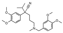
To assess the effects of verapamil on postextrasystolic potentiation (PESP), contrast left ventriculography was performed in ten healthy anesthetized dogs before and after the intravenous administration of verapamil, 0.1 mg/kg. During the contrast ventriculography, a single atrial premature stimulus was introduced. Ejection fractions of a control beat and postpremature beat were measured before and after verapamil. Before verapamil, the mean ejection fractions of the control beat and the postpremature beat were 60 [+ or -] 10 percent and 67 [+ or -] 10 percent, respectively (p<0.05). Following the administration of verapamil, the mean ejection fraction of the control beat decreased from 60 [+ or -] 10 percent to 55 [+ or -] 11 percent (p<0.05). However, the mean ejection fraction of the postpremature beats increased when compared with the control beats following intravenous verapamil (65 [+ or -] 8 percent and 55 [+ or -] 11 percent, respectively; p<0.05). These results suggest that PESP is not inhibited by the administration of intravenous verapamil.
Postextrasystolic potentiation (PESP) is a fundamental property of the mammalian heart.[1,2] The mechanisms responsible for the increased contractility of the postextrasystolic beat are not clear. Increased availability of calcium at the contractile sites is thought to be one of the mechanisms for the PESP.[3] In patients with ischemic heart disease, demonstration of PESP in myocardial segments that show hypokinesis or akinesis at rest is considered evidence of myocardial viability.[4-7] These segments are likely to improve following revascularization. Calcium-channel blockers such as verapamil inhibit the influx of extracellular calcium into the myocardial cell, resulting in negative inotropism.[8] The increased availability of intracellular calcium is believed to be the major mechanism for PESP; therefore, it is logical to hypothesize that PESP may be adversely affected by calcium-channel blocking agents.[9] The present study was designed to investigate the effects of intravenous verapamil on PESP in a canine model.
MATERIALS AND METHODS
Ten adult mongrel dogs weighing 15 to 20 kg were premedicated with acepromazine, 1 mg/kg administered intravenously, and anesthetized with sodium pentobarbital, 20 to 40 mg/kg administered intravenously. Right femoral arterial and venous sheaths were placed by using the Seldinger technique. A 6F bipolar pacing catheter was advanced to the right atrium and placed in a stable position in the high right atrium. An 8F pigtail catheter was advanced to the ascending aorta and subsequently to the left ventricle. Heart rate, mean arterial pressure, and left ventricular end-diastolic pressure were recorded in the control state.
Right atrial pacing was begun at a cycle length 50 ms shorter than the basic sinus cycle length at three times the diastolic threshold by using a model 5325 programmed stimulator (Medtronic, Minneapolis). Left ventricular cineangiography was performed using 45 ml of Renografin-76 (diatrizoate meglumine; E. R. Squibb, New Brunswick, NJ) at a rate of 15 ml/s. During injection of the contrast material, a premature atrial beat was manually induced at a coupling interval that was 100 ms shorter than the basic paced cycle length. After delivery of the premature stimulation, the programmed stimulator reverted to a standby mode, allowing spontaneous sinus rhythm to terminate the postextrasystolic pause.
When the hemodynamic variables returned to baseline following the initial ventriculogram, verapamil, 0.1 mg/kg, was given intravenously over a period of 1 min. Heart rate, mean arterial pressure, and left ventricular end-diastolic pressure were measured 2 min after the administration of verapamil. Atrial pacing was resumed at a cycle length that was 50 ms shorter than the basic cycle length.
Contrast ventriculography was repeated as described previously. A premature atrial stimulus was induced during the ventriculogram at a coupling interval that was 100 ms shorter than the atrial paced cycle length, and sinus rhythm was allowed to resume, terminating the pause following the premature beat.
Left ventricular ejection fractions of a control beat and the postpremature beat, before and after verapamil, were calculated by using the technique described by Yang et al.[10] Statistical analysis was performed with use of the Wilcoxon matched-pairs signed-ranks test to assess the effects of verapamil on hemodynamics and ejection fraction. An a priori level of significance of p<0.05 was used. Data are presented as mean [+ or -] SD.
RESULTS
Technically adequate studies were obtained in eight of the ten dogs. The left ventriculograms of two dogs were not used for analysis because of ventricular arrhythmias during the contrast injection.
The effects of verapamil on hemodynamics are shown in Figure 1. The mean heart rate increased from 117 [+ or -] 18 beats per minute to 130 [+ or -] 14 beats per minute (p<0.05); mean arterial pressure decreased from 95 [+ or -] 12 mm Hg to 82 [+ or -] 10 mm Hg (p<0.05); and left ventricular end-diastolic pressure did not change significantly (p>0.05). Following verapamil administration, the mean ejection fraction of the control beats decreased from 60 [+ or -] 10 percent to 55 [+ or -] 11 percent (p<0.05), and the mean ejection fraction of the postextrasystolic beats decreased from 67 [+ or -] 10 percent to 65 [+ or -] 8 percent (p>0.05) (Fig 2). Before verapamil administration, the mean ejection fractions of the control and postextrasystolic beats were 60 [+ or -] 10 percent and 67 [+ or -] 10 percent, respectively (p<0.05) (Fig 3). Following verapamil, the mean ejection fraction of the postextrasystolic beat was significantly higher compared with control (65 [+ or -] 8 percent and 55 [+ or -] 11 percent, respectively; p<0.05) (Fig 3).
DISCUSSION
Postextrasystolic potentiation is a basic property of the myocardium and has been used in the evaluation of contractile reserve. In the intact human heart, at least three mechanisms appear to play important roles in PESP:[3] (1) a decrease in aortic impedance, because the compensatory pause provides time for increased peripheral runoff; (2) increased diastolic filling of the left ventricle during the compensatory pause, which results in increased preload and hence contractility related to the Frank-Starling mechanism; and (3) increased availability of calcium at the contractile sites.
Because PESP is observed in isolated isometrically contracting muscle, it has been suggested that increased availability of intracellular calcium is probably the major mechanism for the potentiation.[11] Calcium-channel blocking agents, which are widely used in patients with ischemic heart disease, produce significant cardiovascular effects by inhibiting the influx of calcium across the cell membrane.[8,12] These effects include systemic and coronary vasodilation, depression of myocardial contractility, and a decrease in atrioventricular conduction. Since PESP is believed to be at least partly mediated through increased availability of calcium ions, it is possible that calcium-channel blocking agents could prevent or modify PESP. Our study suggests that PESP is not significantly altered by calcium-channel blockers in the intact healthy dog.
The decreased ejection fraction following verapamil administration, in spite of an increase in heart rate and a decrease in mean arterial pressure, suggests that negative inotropic effects were induced by administration of the drug. The lack of any significant effect of verapamil on PESP may be due to several factors. The systemic vasodilation and increased sympathetic stimulation caused by verapamil might have negated any negative inotropic effects. However, this is unlikely because the ejection fraction of the control beats decreased following the administration of verapamil. The compensatory pause following premature stimulation resulted in a significant decrease in afterload and an increase in preload and thereby increased the ejection fraction. Although it has been previously shown that PESP can be demonstrated in beats without a compensatory pause,[14] we cannot exclude this mechanism as the cause of the potentiation. It is possible that higher doses of intravenous verapamil might have inhibited PESP. However, we selected a dose of 0.1 mg/kg for this study on the basis of previous studies,[15] which have shown that a significant negative inotropic effect results from intravenously administering this dose. Finally, we cannot exclude the possibility that other, as yet unidentified mechanisms may be responsible for PESP.
There are several potential limitations in this study. The use of ejection phase indices for assessment of inotropic function is problematic. Use of the end-systolic volume-stress relationship would have been more reflective of inotropic function. The duration of the compensatory pause following the premature beat at baseline and that following verapamil were not controlled. However, the compensatory pause following verapamil was actually shorter because of the increase in spontaneous sinus rate. We did not measure left ventricular volumes of the pre-and postpremature beats. Ejection fraction was calculated by using a simplified formula incorporating end-diastolic and end-systolic area measured by planimetry.[11] Lack of volume data does not negate the validity of ejection fraction measurement. Atrial premature stimulation was used instead of ventricular stimulation in order to ensure atrioventricular synchrony during the pre-and postpremature beats. Previous studies have shown that atrial stimulation can be used in the assessment of PESP.[16]
In conclusion, in spite of the inherent limitations of extrapolating from animal experimental data to the clinical situation, our study suggests that calcium-channel blockers do not alter PESP.
ACKNOWLEDGMENTS: We would like to acknowledge the technical assistance of Richard Mathias and the secretarial assistance of Ann Clark.
REFERENCES
1 Langendorff O. Uber elektrishe Reizung des Herzens. Arch Anat Phys Abst 1885; 284
2 Hoffman BF, Bindler E, Suckling EE. Postextrasystolic potentiation of contraction in cardiac muscle. Am J Physiol 1956; 185:95
3 Cohn PF. Evaluation of inotropic contractile reserve in ischemic heart disease using postextrasystolic potentiation. Circulation 1980; 61:1071-75
4 Hamby RI, Aintablian A, Wisoff G, Hartstein ML. Response of the left ventricle in coronary artery disease to post extrasystolic potentiation. Circulation 1985; 51:428-35
5 Popio KA, Gorlin R, Bechtel D, Levine JA. Postextrasystolic potentiation as a predictor of potential myocardial viability: preoperative analyses compared with studies after coronary artery bypass surgery. Am J Cardiol 1977; 39:944-53
6 Cohn PF, Gorlin R, Herman M, et al. Relation between contractile reserve and prognosis in patients with coronary artery disease and depressed ejection fraction. Circulation 1975; 51:414-20
7 Cohn LH, Collins JJ, Cohn PF. Use of the augumented ejection fraction to select patients with left ventricular dysfunction for coronary revascularization. J Thorac Cardiovasc Surg 1976; 72:835-39
8 Fabiato A, Fabiato F. Calcium and cardiac excitation contraction coupling. Ann Rev Physiol 1979; 41:473
9 Ilia R, Rudnik L, Gueron M. Lack of postextrasystolic potentiation in a normal heart. Cathet Cardiovasc Diagn 1987; 13:189-90
10 Yang, Bentivoglio, Marachao, Goldberg. Cardiac catheterization data to hemodynamic parameters. Philadelphia: FA Davis, 1978; 89:151
11 Cohn PF. Contractile reserve in coronary heart disease: detection and clinical importance. Prim Cardio 1981; 7:48-53
12 Nayler WG, Merrillees NCR. Cellular exchange of calcium. In: Harris, Lop, eds. Calcium and the heart. New York: Academic Press, 1971; 24-65
13 Adam RJ, Schwartz A. Comparative mechanisms for contraction of cardiac and skeletal muscle. Chest 1980; 78:123-39
14 Kuijer PJ, van der Werf T, Meijler FL. Post-extrasystolic potentiation without a compensatory pause in normal and diseased hearts. Br Heart J 1990; 63:284-86
15 Walsh RA, Badke FR, O'Kourke RA. Differential effects of systemic and intracoronary calcium channel blocking agents on global and regional left ventricular functions in conscious dogs. Am Heart J 1981; 102:341-50
16 Kalff V, Chan W, Rabinovitch M, O'Neill W, Walton J, Steward J, et al. Radionuclide evaluation of postextrasystolic potentiation of left ventricular function induced by atrial and ventricular stimulation. Am J Cardiol 1982; 50:106-11
COPYRIGHT 1992 American College of Chest Physicians
COPYRIGHT 2004 Gale Group


