What you need to know about diagnosing and treating viruses.
The cornea is a unique tissue. Capable of only a limited range of responses to a wide variety of insults, its differential diagnosis of infection can be complex and perilous. Delay or misdiagnosis can be disastrous.
Viral infection of the cornea is common; yet, despite the limited number of potential pathogens, the spectrum of infection is broad and the clinical presentation often confusing.
We recently saw a patient who was a perfect example of the viral keratitis diagnosis dilemma.
Misled by Maria
Even the experienced clinician can be misled. Take the case of Maria, a 56-year-old woman who presented to us with discomfort in her left eye.
Because Maria didn't speak English, getting a complete history was difficult. After some investigation, it seemed that she'd been treated for a corneal ulcer 2 weeks previously at a local medical center. Maria was taking ciprofloxacin hydrochloride (Ciloxan) q.i.d., and strangely (for it delays healing), fluorometholone (FML) t.i.d. OS.
Best corrected visual acuity was 20/25- OS, with no improvement through a pinhole occluder. Slit lamp evaluation revealed a small, circumscribed, round lesion with an overlying punctate keratitis. The anterior chamber was deep and quiet.
We diagnosed a resolving corneal ulcer, and discontinued the fluorometholone. We told Maria to return the next day.
She returned a week later reporting improvement but still complaining of mild discomfort in the left eye.
Visual acuity was still 20/25 OD and 20/40 OS. Slit lamp evaluation revealed the same stromal lesion with mild overlying punctate staining, but there was no rose bengal staining.
We noted trace cells in the anterior chamber OS. Intraocular pressures measured 20 mm Hg OD and 15 mm Hg OS.
Because Maria was feeling better, we again diagnosed a resolving corneal ulcer.
We told her to continue ciprofloxacin q.i.d. and added homatropine hydrobromide (Homatropine) 5% for the anterior chamber reaction and her comfort. We asked her to return in 2 days. Again, she came a week later, this time with her son as interpreter.
No improvement
She reported that her left eye felt much worse and her eyesight had also worsened. Her son said that the problems with his mother's eye had recurred several times over the past 20 years.
Visual acuity was 20/60 OS, with no improvement through a pinhole. Slit lamp evaluation revealed significant corneal haze surrounding a mildly excavated round lesion, which stained with sodium fluorescein. We applied rose bengal again and saw staining around the edges of the lesion, indicating devitalized cells.
Upon testing, corneal sensitivity was reduced. The anterior chamber was deep and quiet.
We diagnosed a recurrent herpes simplex keratitis, discontinued ciprofloxacin and homatropine, and placed her on trifluridine (Viroptic Ophthalmic Solution 1%) q2h for 48 hours, after which she was to return.
Feeling better
Maria returned 3 days later feeling much better. She reported only slight discomfort and blurred vision. Visual acuity was still 20/60 OS. Slit lamp evaluation revealed slight corneal haze with no rose bengal staining.
On closer evaluation, we saw pooling of fluorescein over an area of stromal loss. We determined this by gently applying a cotton swab to the cornea. Any residual fluorescein was absorbed, leaving nothing but the depression. Had there been an epithelial break, fluorescein would have remained in the stroma and continued to stain.
The stromal herpetic keratitis was resolving. We kept Maria on trifluridine 5 times a day and prednisolone acetate (Pred-Forte) q.i.d. because there was no longer an epithelial defect.
Maria returned 1 week later complaining of a scratchy sensation OS upon awakening.
Her visual acuity was 20/25 OD and 20/30 OS. Very mild stromal haze remained around the resolving scar, with no staining. We began slowly tapering the steroids (t.i.d. x 1 month, b.i.d. x 1 month, qd x 1 month and q.o.d. x 1 month) while maintaining the antiviral coverage t.i.d. Once we were down to every other day, we discontinued the trifluridine.
Maria returned to us monthly for follow-ups and intraocular pressure checks. Each time she said she felt better, and she reported no discomfort or blurred vision. However, a small stromal scar remained.
It's easy to see how initially, without a thorough history, we were misled. If we had realized sooner that Maria had herpetic keratitis, quicker management with trifluridine may have prevented stromal infection, and probably scarring too.
Viruses you'll need to know
Viruses that affect the cornea can range from mild and selflimiting to uncontrolled and sight-threatening. The viruses that most commonly affect the cornea are herpes simplex virus (HSV), varicella zoster virus and adenoviruses.
Herpes simplex virus. HSV is a member of the herpes virus group, which also includes varicella zoster, Epstein-Barr and cytomegaloviruses. It occurs in two immunologic forms, Type 1 infecting the mouth or nares, and Type II causing genital herpes.
Most corneal infections and fever blisters are caused by HSV I. The virus is latent in the ganglia and waits for a recurrence of compromise in the host's immune system. Most recurrences happen during the first year after the initial attack.
Man is the only known host of HSV, which spreads only through direct contact. Herpes simplex keratitis is the leading cause of blindness in the United States, with nearly 500,000 cases reported annually.
Epithelial keratitis. Epithelial keratitis I is caused by virus replicating within the epithelium, resulting in cell death.
The presentation is typically unilateral with foreign body sensation, photophobia, tearing and reduced acuity. Occasionally, symptoms can be mild because of HSV-induced hypesthesia of the cornea.
Corneal ulcers can begin as punctate keratitis, which usually but not always coalesces to form the arborizing dendrites pathognomonic to HSV. The virus replicates most intensely in the cells at the lesion's edges. Complete epithelial loss exposes Bowman's membrane at the ulcer's center
Don't use steroids indiscriminately because this can enhance progression of the keratitis to disciform keratitis and geographic ulceration.
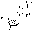
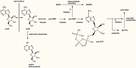
Treatment. Debridement and antiviral agents are the mainstays of epithelial HSV keratitis treatment. For best results, use trifluridine drops q2h for the first 48 hours with vidarabine 3% (Vira-A) every hour at night. Trifluridine may be tapered to 5 to 9 times as the lesions resolve.
Never use steroids while an epithelial defect remains. Once the epithelium has been intact with a few days of antiviral coverage, consider steroids to reduce corneal scarring. Prednisolone acetate q.i.d. is usually sufficient and should be tapered slowly
The use of oral acyclovir in treating stromal keratitis is controversial, but recent studies show no statistically or clinically significant benefits in treating HSV stromal keratitis in patients receiving concomitant topical corticosteroids and trifluridine.
* Varicella zoster virus. This virus, also known as shingles (it causes chickenpox as well), is a painful vesicular skin rash. When the virus affects the first division of the trigeminal nerve, the resulting disease is termed herpes zoster ophthalmicus. As with HSV, zoster establishes latency in the ganglia and waits to recur.
Immunodeficiency predisposes a patient to recurrence, and human immunodeficiency virus (HIV) should be ruled out if its risk factors are evident.
Ophthalmic complications occur when the nasociliary branch of V1 is affected. This nerve also innervates the tip of the nose through its terminal branch, the ethmoidal nerve.
Vesicles on the end of the nose (Hutchinson's sign) suggest more severe ocular involvement such as conjunctivitis, epithelial and stromal keratitis, tear film dysfunction, epithelial defects, neurotrophic keratopathy, stromal melting and band keratopathy
Early punctate staining can be self-limiting and is managed only with frequent lubrication.
For conjunctivitis, steroidantibiotic combinations help. Topical steroids reduce stromal inflammation and scarring, and anterior uveitis.
Steroids aren't contraindicated for epithelial zoster lesions as they are for HSV. Oral acyclovir within a few days of onset reduces subsequent ocular complications and number and duration of subsequent eruptions.
Adenovirus. Dozens of adenoviruses exist, but the most common are types 8 and 19, associated with epidemic keratoconjunctivitis (EKC), and types 3, 4, 7 and 14, associated with pharyngoconjunctivitis. Hardy and contagious, they can remain potentially infectious for at least a week on surfaces in your office, especially the tonometer tip.
EKC. This condition usually presents with a unilateral painful, tearing eye. The second eye is usually affected within a week. Clinical signs, which are self-limiting, are papillary or follicular conjunctivitis, chemosis, serous discharge, preauricular adenopathy and mild uveitis.
Pseudomembranous or subconjunctival hemorrhages may develop. Keratitis usually begins around the third week, forming the characteristic subepithelial infiltrates that can decrease acuity if found centrally
There's no effective treatment for EKC. Supportive therapy of cold compresses, lubricants, nonsteroidal anti-inflammatory drugs (NSAIDs) and vasoconstrictors may help.
Steroid use is controversial; reserve them for severe cases. Steroids increase comfort but don't speed healing and may take months to taper. Infiltrates may reappear, sometimes worse than before.
Pharyngeal conjunctival fever (PCF ). PCF is acute pharyngitis with follicular conjunctivitis and low-grade fever. It's most common in children and presents with acute red, puffy, watery eyes with foreign body sensation.
You'll typically find a preauricular node. The patient's other eye is usually affected too, but less severely.
Unlike EKC, PCF rarely produces significant infiltrates or long-term ocular complications. Symptoms usually last 1 to 3 weeks; therapy is symptomatic.
Proceed carefully
A complete case history and thorough examination are necessary for correct diagnosis and therapy. Remain focused, follow the patient closely until you see improvement, and realize when it's in the patient's best interest to refer to an ophthalmologist. OM
Dr. Morschauser is in private practice at the New York Eye and Ear Infirmary in New York City. Dr. Metwalli is on staff at Duke University Hospital in the Comprehensive Ophthalmology Department.
Copyright Boucher Communications, Inc. Dec 1998
Provided by ProQuest Information and Learning Company. All rights Reserved


