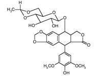
Thirty-three patients with T3,N2,M0 or T4,N2,M0, nonsmall-cell lung cancer (NSCLC) took part in a phase 2 study in an attempt to evaluate the feasability of neoadjuvant chemotherapy followed by surgery and thoracic radiotherapy. Chemotherapy consisted of daily administration of the following treatment: etoposide, 100 mg/[m.sup.2]; cisplatin, 25 mg/[m.sup.2]; ifosfamide, 1.5 g/[m.sup.2]; and mesna, 1.8 g/[m.sup.2] for 4 days. Three cycles were planned starting every 21 days. Responding patients underwent a thoracotomy in order to attempt a resection and then received a 45 Gy of thoracic radiotherapy. The results of response and resection rates have been published and the present final report deals with the longterm results. Chemotherapy induced a 55 percent partial response rate and a 15 percent complete response rate allowing a complete resection in 55 percent of the patients. Complete remission was histologically confirmed for the five complete responders. Although the median survival was short (10 months), six patients were long-term survivors (3-year survival rate: 19 percent). Survival was significantly influenced by the type of resection: patients for whom a complete resection was possible survived the longest with a median survival three times that of the other patients. Modalities of relapses differed according to the results of surgery: 8 of the 15 patients who did not undergo a complete surgical resection experienced a local relapse during the first 18 months of follow-up whereas in the complete resection group, central nervous system metastasis was the main site of relapse. We conclude that the neoadjuvants ifosfamide, cisplatin, and etoposide in patients with locally advanced NSCLC are feasible to use and allow a 19 percent 3-year survival rate. These results are the rationale of an ongoing randomized study comparing neoadjuvant chemotherapy followed by surgery and surgery alone. This study is designed to test whether neoadjuvant chemotherapy improves survival of patients with locally advanced NSCLC.
(Chest 1994; 106:1451-55)
NSCLC=non-small-cell lung cancer;
TNM=tumor-nodemetastasis;
UICC=International Union Against Cancer;
WHO=World Health Organization.
Key words: feasibility; neoadjuvant chemotherapy; non-small-cell lung cancer (NSCLC); surgery; TNM classification
Non-small-cell lung cancer (NSCLC) is frequently associated with a metastatic disease at some time during the microscopic stage rather than when it becomes clinically detectable.(1) Patients with a locally advanced NSCLC, particularly stage N2, are sometimes considered for surgery, but resection is frequently followed by metastatic relapses.(2) There are two underlying problems where local and regional extension in NSCLC is concerned: first, this extension limits the possibility for a surgeon to carry out a complete resection; second, local extension makes the existence of a microscopic metastatic disease predictable. These clinical observations are the rationale for the evaluation of neoadjuvant chemotherapy as an experimental approach.(3)
In 1987, we started a phase 2 trial of neoadjuvant ifosfamide, cisplatin, and etoposide in patients with locally advanced NSCLC. The response rate, resection rate, and microscopic findings of this study have been published.(4) The present report deals with modalities of failures and long-term results.
METHODS
The method of this study has been previously described in detail.(4) Briefly, patients of both sexes with locally advanced and histologically proven NSCLC were entered into the study. Prerequesites for inclusion were as follows: age <75 years; World Health Organization (WHO) performance status [less than or equal to]2; no distant metastasis; weight loss [less than or equal to!10 percent; respiratory function compatible with surgical resection; normal baseline renal and cardiac functions; baseline neutrophil count [greater than or equal to]2,000/[micro]L; and platelet count [greater than or equal to]100,000/[micro]L. None of them had received prior therapy. For all patients, staging procedure included clinical examination, chest radiographs, computed tomographic (CT) scan of chest, upper abdomen, and brain, fiberoptic bronchoscopy, and bone scanning. Staging was carried out according to the fourth edition of the UICC TNM classification.(5)(6) Patients with gross mediastinal involvement, ie, more than two ipsilateral nodal stations with mediastinal lymph nodes greater than 20 mm on CT scan, and widened carina or main bronchus distortion suggestive of subcarenal lymph nodes, were considered as having locally advanced NSCLC and were included directly in the study; patients for whom this staging procedure failed to demonstrate bulky mediastinal involvement had cervical mediastinoscopy.
Chemotherapy consisted of daily administration of the following treatment: etoposide, 100 mg/[m.sup.2]; cisplatin, 25 mb/[m.sup.2]; ifosfamide, 1.5 g/[m.sup.2]; and mesna, 1.8 g/[m.sup.2] for 4 days. A cycle started every 21 days. Patients were evaluated for response at the start of each cycle of chemotherapy by clinical examination and chest radiograph. Based on the response after the second cycle of chemotherapy, patients with evidence of major response underwent a new complete staging procedure to evaluate it precisely and a resection was planned at that time, whereas other patients underwent a third cycle before complete staging procedure. Chemotherapy-induced tumor response was defined according to WHO:(7) a complete response was defined as the complete disappearance of all lesions; a partial response was defined as equal to or greater than a 50 percent reduction in the product of the two longest perpendicular diameters of the indicator lesions measured on CT scan. For patients who achieved a complete or a partial response, a thoracotomy was scheduled 2 weeks after hematologic recovery with an attempt at curative resection and mediastinal lymph node disection. A complete resection was defined as resection of all macroscopic disease and normal histologic features of the margin. Patients with stable or progressive disease were not planned for surgery. All patients received thoracic radiotherapy (45 Gy, postoperatively or as unique local treatment).
A [x.sup.2] test was used for comparison of frequency distribution of qualitative variables. Survival was defined as the time from the first day of treatment to the date of death. Time to progression was defined as the time from the first course of treatment to the time of relapse for responding patients or progression for patients with stable disease. Probability of survival was estimated by the Kaplan-Meier method.(8) Univariate analysis of survival was done by log-rank test.(9)
RESULTS
Patients
Between September 1987 and September 1989, 33 patients took part in the study. All patients had measurable pathologically confirmed NSCLC. Among them were 22 squamous cell carcinomas, 5 adenocarcinomas, and 6 large-cell carcinomas. For all patients, staging procedure disclosed gross mediastinal lymph node involvement. Invasive staging techniques were needed in 10 of the 33 patients. Thirty-two patients were at stage T3,N2,M0 and one was at stage T4, N2, M0.
Response
Objective responses to ifosfamide, cisplatin, etoposide chemotherapy were obtained in 23 of 33 patients. Among them, a complete response was achieved in 5 patients (15 percent) and a partial response was achieved in 18 patients (55 percent). Stable disease was observed in three patients and progressive disease occurred in seven patients. Progression during the chemotherapy program consisted of metastases in five patients and local progression in the other two.
Toxicity
The main toxic reaction of ifosfamide, cisplatin, and etoposide chemotherapy was a moderate to severe hematologic one. Ninety-two percent of the patients experienced a grade 3 to 4 neutropenia toxic reaction during the chemotherapy program and 60 percent experienced a grade 3 to 4 thrombopenia (according to the WHO scale). Among them, 10 patients developed a grade 2 to 3 infection. These patients required a hospitalization lasting for 6 days and intravenous antibiotics. Blood transfusions were given to six patients and platelet transfusions were given to four others. One patient died of central nervous system hemorrhage.
Surgery and Microscopic Findings
The results of surgery and pathologic examination of the specimen are shown in Table 1. The overall complete resection rate was 55 percent (18/33). Among them, no macroscopic or microscopic remainders were seen for the five complete responders. Three additional patients were operated on: one open and close and two incomplete resections. No surgery was envisaged for the remaining 12 patients owing to restaging procedure demonstrating an inoperable disease. No additional morbidity was observed after surgery. The frequency of a complete resection did not significantly differ when patients with a prestudy positive lymph node biopsy specimen were compared with patients for whom lymph node involvement had been demonstrated by CT scan only (66 percent vs 50 percent; [x.sup.2]: 0.73; NS).
Table 1--Resection Rate and negative Histology Following Neoadjuvant
VIP(*)
(*)VIP = ifosfamide, cisplatin, etoposide combination; CR = complete response; PR = partial response; SD = stable disease; PD = progressive disease.
Survival
Median follow-up duration was 16 months (range, 1 to 64 months). One patient was unavailable for follow-up. Median survival was 10 months. Probabilities of survival at 1, 2, and 3 years were 43, 28, and 19 percent, respectively (Fig 1). In responding and stable patients, median time to progression was 16.7 months (Fig 2). Patients who underwent a complete resection proved to have a significantly longer time to progression and overall survival when compared with patients who underwent either incomplete or no resection (median survival, 20.1 and 6.7 months for the former and latter patient subgroups, respectively; log-rank test: p<0.0001; Fig 3). Histologic subtype, ie, squamous vs others, grade III to IV, hematologic toxicity, and pretreatment weight loss had no significant effect on time to progression and overall survival. The survival of patients who underwent a prestudy mediastinoscopy demonstrating pathologic N2 did not significantly differ when compared with patients for whom only CT scan was carried out to demonstrate stage N2 (median survival, 15.4 and 10 months in the former and the later subgroup, respectively).
[CHART OMITTED]
Patterns of Relapses
Relapse patterns were studied separately according to the type of resection (Table 2). Among the 18 patients who underwent a complete resection, 6 were alive at time of reporting, 4 died of intercurrent diseases (2 pulmonary embolisms and 2 myocardial infractions), and 8 experienced a relapse. For these latter patients, distant metastasis was the main modality of relapse with central nervous system representing the unique site in four.
Table 2--Modalities of Relapse According to Resection Type
(*)One patient with incomplete resection was unavailable for followup.
Among the 15 patients for whom resection was either incomplete or not done, none survived more that 18 months after the induction therapy, one died after cycle 1 of central nervous system hemorrage, and 13 experienced a relapse. Local progression was the main modality of relapse for those patients (8/15). Conversely, brain metastasis seems to be a rare event in this subgroup as only one patient had this modality of relapse. All six long-term survival patients have had a complete resection and are free fromdisease at time of reporting (Table 3). Three of them have had biopsy-proven stage-N2 found by prestudy mediastinoscopy. Half of the long-term survivors responded completely to chemotherapy and the others were partial responders. In all cases, results of the pathologic examination of the resection showed normal nodal status.
DISCUSSION
This study shows that neoadjuvant ifosfamide, cisplatin, and etoposide is an active combination in patients with locally advanced NSCLC inasmuch as it induced complete pathologic response in 15 percent of the patients and allowed a complete resection in 55 percent. This antitumor activity is associated with a 19 percent 3-year survival. The modality of relapse differs when patients who underwent a complete resection are compared with others, the central nervous system remaining the first site of failure in the former group.
Neoadjuvant chemotherapy was tested in patients with NSCLC in an attempt to increase the resectability of the tumor and to treat the microscopic metastatic disease known to be responsible for the majority of failures in surgically treated patients. Several published trials have been reviewed extensively.(1)(9)(10) Most of them are feasibility studies in stage III NSCLC. Obviously, the heterogeneity of eligibility criteria from one study to another prevents general conclusions on the usefulness of neoadjuvant chemotherapy. In addition, the majority of the studies, including the one reported herein, have been designed as phase 2 trials and, therefore, they cannot answer the following questions. Is the operability of patients with NSCLC increased by neoadjuvant chemotherapy? Is overall survival improved by this approach? However, it is possible to conclude that neoadjuvant chemotherapy has an antitumor activity: most studies report a 60 percent objective response rate, including a significant number of complete responses and a 50 percent complete resection rate. Neoadjuvant chemotherapy does not increase morbidity after surgery except when it is combined with preoperative radiation therapy.(1)(10)
The results of our study show similar rates of chemotherapy-induced tumor responses, complete pathologic responses, and complete resections. The staging procedure we used could be discussed. In the literature, some studies used a radiographic definition of mediastinal lymph node enlargement (clinical N2)(11)(12) whereas others used a strict pathologic definition of this involvement.(13)(14) Although a real comparison of these two groups of studies is not possible for methodologic reasons, there is no major difference when resection rates and survivals are considered. In our study, we decided to avoid mediastinoscopy for patients with bulky N2 on CT scan and to determine N2 disease pathologically in the other cases. We observed no difference in resection rate and survival when these two groups were compared. In addition, three of six of the long-term survival patients had a biopsy-proven N2 stage tumor at time of inclusion.
The analysis of survival in our study showed that long-term survival can be achieved in patients with N2 disease receiving neoadjuvant chemotherapy. Median survival was calculated as 10 months by the Kaplan-Meier method and suggested a weak advantage of neoadjuvant ifosfamide, cisplatin, and etoposide. However, the median survival is only a rough indicator of prognosis, particularly in a small size population and the 3-year survival rate might be a better variable to evaluate a study. The 19 percent 3-year survival observed in our study can be regarded as sufficiently favorable to consider our protocol as feasible. A clear survival advantage has been observed for patients who underwent complete resection. However, it is not possible to assume that surgical excision is the only reason for such a result and some other factors, including response to chemotherapy, might be considered.
In the published studies, median survival varied from 8 to 24 months. This wide range might be explained by (1) true differences in efficacy between protocols, and (2) heterogeneity of eligibility criteria. When available, long-term survival reports show that a 15 percent 3-year survival rate can be expected for patients who benefit from neoadjuvant chemotherapy. These results are not a proof of the chemotherapy efficacy but deserve further randomized studies designed to test the effectiveness of neadjuvant chemotherapy in NSCLC. In this setting, a possible study design to determine the survival advantage of neoadjuvant chemotherapy might be to randomize patients with stage N2 NSCLC between neoadjuvant chemotherapy followed by surgery and surgery alone. A recent study investigating this comparison concluded that neoadjuvant chemotherapy increases the median survival of stage IIIa NSCLC.(15)
In March 1990, we started a randomized phase 3 trial based on a similar design. The aim of this study is to determine whether enhanced response and resection rates could increase survival of patients with NSCLC treated by ifosfamide, cisplatin, and etoposide neoadjuvant chemotherapy. Other trials based on alternative methods of randomization are also ongoing and one could expect that these studies could define the place for neoadjuvant chemotherapy in NSCLC.
ACKNOWLEDGMENT: We are indebted to Dr. C. Marty-Ane, Dr. V. Gautier, Dr. R. Arriagada, Dr. D. Grunenwald, and Pr. P. Godard for their cooperation; and to Mrs. Jo Baissus for help in preparing the manuscript.
REFERENCES
(1)Shepherd FA. Induction chemotherapy for locally advanced non-small cell lung cancer. Ann Thorac Surg 1993; 55:1585-92
(2)Naruke T, Suemasu K, Ishikawa S. Lymph node mapping and curability at various levels of metastasis in resected lung cancer. J Thorac Cardiovasc Surg 1978; 76:832-39
(3)DeVita VT. On the value of response criteria in therapeutic research. Bull Cancer 1988; 75:863-69
(4)Pujol JL, Rossi JF, Le Chevalier T, Daures JP, Rouanet P, Douillard JY, et al. Phase II pilot study of neoadjuvant ifosfamide, cisplatin and etoposide in locally advanced non-small cell lung cancer. Eur J Cancer 1990; 7:798-801
(5)Tisi GM, Friedman PJ, Peters RM, Pearson G, Carr D, Lee RE, et al. American Thoracic Society: clinical staging of primary lung cancer. Am Rev Respir Dis 1982; 125:659-64
(6)Mountain CF. A new international staging system for lung cancer. Chest 1986; 89(suppl4):225S-33S
(7)World Health Organization. WHO handbook for reporting the results of cancer treatment. Geneva: WHO Offset Publication No. 48, 1979:1-35
(8)Kaplan EL, Meier P.Nonparametric estimation from incomplete observations. J Am Stat Assoc 1958; 53:457-81
(9)Murren JR, Buzaid AC, Hait WN. Critical analysis of neoadjuvant therapy for state IIIa non-small cell lung cancer. Am Rev Respir Dis 1991; 143:889-94
(10)Pujol JL, Le Chevalier T, Ray P, Gautier V, Grunewald D, Miche FB. Neoadjuvant chemotherapy of locally advanced non-small cell lung cancer. Lung Cancer (in press)
(11)Martini N, Kris MG, Flehinger BJ, Gralla RJ, Bains MS, Burt ME, et al. Preoperative chemotherapy for stage IIIa (N2) lung cancer: the Sloan-Kettering experience with 136 patients. Ann Thorac Surg 1993; 55:1365-74
(12)Taylor SG, Trybula M, Bonomi PD, Faber LP, Lee MS, Reddy S, et al. Simultaneous cisplatin fluorouracil infusion and radiation followed by surgical resection in regionally localized stage III, non-small cell lung cancer. Ann Thorac Surg 1987; 43:87-91
(13)Strauss GM, Herndon JE, Sherman DD, Mathisen DJ, Carey RW, Choi NC, et al. Neoadjuvant chemotherapy and radiotherapy followed by surgery in stage IIIA non-small cell carcinoma of the lung: report of a cancer and leukemia group B phase II study. J Clin Oncol 1992; 10:1237-44
(14)Vokes EE, Bitran JD, Hoffman PC, Ferguson MK, Weichselbaum RR, et al. Neoadjuvant vindesine, etoposide, and cisplatin for locally advanced non-small cell lung cancer: final report of a phase II study. Chest 1989; 96:110-13
(15)Rosell R, Gomez-Codina J, Camps C, Maestre J, Padille J, Canto A, et al. A randomized trial coparing preoperative chemotherapy plus surgery with surgery alone in patients with non-small cell lung cancer. N Engl J Med 1994; 330:153-58
COPYRIGHT 1994 American College of Chest Physicians
COPYRIGHT 2004 Gale Group


