Tremor--a rhythmic, involuntary, oscillatory movement of body parts (1)--is the most common movement disorder. (2) The diagnosis is based on a careful assessment of the history and physical examination, although some tests, including positron emission tomography (PET) and single photon emission computed tomography (SPECT), are being investigated as diagnostic aids. (2-5) This article reviews the classification and causes of tremor and provides evaluation guidelines.
Classification
Tremors are classified as rest or action tremors. Rest tremor occurs when the affected body part is completely supported against gravity (e.g., hands resting in the lap). Amplitude increases during mental stress (e.g., counting backwards) or with general movement (e.g., walking) and diminishes with target-directed movement (e.g., finger-to-nose test). (1,2,6)
Action tremors are produced by voluntary muscle contraction. They are further divided into postural, isometric, or kinetic tremors. Postural tremor occurs when the affected body part maintains position against gravity (e.g., extending arms in front of body). Isometric tremor results from muscle contraction against stationary objects (e.g., squeezing the examiner's fingers). Kinetic tremor, which occurs with voluntary movement, is either simple kinetic tremor or intention tremor. Simple kinetic tremor is associated with movement of extremities (e.g., pronation-supination or flexion-extension wrist movements). Intention tremor occurs during visually guided movement toward a target (e.g., finger-to-nose or finger-to-finger testing), with significant amplitude fluctuation on approaching the target (2) (Table 1). (1,6)
Although this classification helps in determining cause, the presentation of tremor syndromes varies. Other aspects of the history and physical examination should be considered when evaluating patients with tremor.
Tremor Syndromes
PHYSIOLOGIC TREMOR
All normal persons exhibit physiologic tremor, a benign, high-frequency, low-amplitude postural tremor. Usually invisible to the naked eye, it can be amplified by holding a piece of paper on the outstretched hand or pointing a laser at a distant screen. (1,2)
Enhanced physiologic tremor is a visible, high-frequency postural tremor that occurs in the absence of neurologic disease and is caused by medical conditions such as thyrotoxicosis, hypoglycemia, the use of certain drugs, or withdrawal from alcohol or benzodiazepines. It is usually reversible once the cause is corrected1,2 (Table 2). (6,7)
ESSENTIAL TREMOR
Essential tremor is a visible postural tremor of hands and forearms that may include a kinetic component. (1) It is the most common movement disorder worldwide; prevalence ranges from 4.1 to 39.2 cases per 1,000 persons, to as high as 50.5 per 1,000 in persons older than 60 years. (8) These figures may underestimate the true prevalence, however, because up to 50 percent of persons with mild essential tremor are unaware of it. (9) Reports of family history vary widely, with 21.7 percent of patients in one study (9) and 62 percent in another study (10) reporting a family history of tremor.
Essential tremor develops insidiously and progresses slowly, presenting as a postural, distal arm tremor in 95 percent of patients. Onset peaks bimodally in the teens and 50s. The tremor may start in a single limb, but it becomes bilateral over time, most often as a flexion-extension movement of the wrist with a frequency of 4 to 12 Hz. It may involve the head, appearing as a yes-yes or no-no head movement. Amplitude increases with stress, fatigue, and certain medications such as central nervous system stimulants, and may increase with certain voluntary activities such as holding a fork or cup. Rest, beta blockers, primidone (Mysoline), and alcohol ingestion decrease the tremor. (2,10,11)
PARKINSON'S DISEASE
Parkinson's disease (PD) is 20 times less common than essential tremor. (8) Nevertheless, approximately 1 million Americans have PD. (12) Because specific treatment options are available, accurate diagnosis is essential. (2,6,12,13)
Symptoms develop insidiously, often after age 50, although early-onset disease may appear in the 20s. (13) Initial symptoms include resting tremor beginning distally in one arm at a 4- to 6-Hz frequency. Typically, the tremor is a flexion-extension elbow movement, a pronation-supination of the forearm, or a pill-rolling finger movement. It worsens with stress and diminishes with voluntary movement. It may have postural or kinetic components. (2,12) However, 10 to 20 percent of patients have no tremor during the course of PD. (2,14)
Other signs of PD include rigidity, bradykinesia, and impaired postural reflexes. The physician may note cogwheel rigidity (i.e., ratchet-like resistance) during passive range of motion while examining the extremities. Bradykinesia includes a slow, shuffling gait, decreased arm swing with walking, difficulty rising from a seated position, and reduced facial animation (masked facies). (12,13,15) Postural reflexes are examined by the pull test: the patient stands with arms hanging loosely at the sides; from behind, the examiner holds the patient's upper arms just under the shoulders and gently pulls backward; if the patient begins to fall, postural instability is indicated (12) (Table 3). (16)
CEREBELLAR TREMOR
Cerebellar tremor presents as a unilateral or bilateral, low-frequency (less then 5 Hz) intention tremor caused by stroke, brainstem tumor, or multiple sclerosis. (2,17) It may include postural tremor. (1) Classically, cerebellar lesions produce kinetic tremor on the ipsilateral side of the body. Finger-to-nose, finger-to-finger, and heel-to-shin testing results in worsening tremor as the extremity approaches the target. (2) Other signs include abnormalities of gait, speech, and ocular movements; inability to perform rapid alternating hand movements; (6) and titubation, a postural tremor of the trunk and head. (5)
DRUG-INDUCED AND TOXIC TREMORS
Drug-induced tremor may follow ingestion of certain drugs (Table 4) (1); toxic tremors occur following intoxication. Tremors also are present during withdrawal from certain drugs and alcohol.1
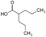
The most common drug-induced tremor is enhanced physiologic tremor following use of sympathomimetics such as pseudoephedrine, bronchodilators, or theophylline, and antidepressants such as tricyclics or fluoxetine (Prozac). (1,7) This tremor also may accompany benzodiazepine withdrawal. (2,7) Approximately 25 percent of patients taking long-term valproic acid (Depakene) therapy exhibit postural tremor three to 12 months after starting therapy. Lowering the dosage decreases the tremor. (2)
Lithium can induce a fine postural tremor of the hands (8 to 12 Hz). Directly correlated with serum concentration, lithium toxicity may cause permanent damage to the cerebellum that precipitates postural and intention tremors. Amiodarone (Cordarone) may cause a dose-dependent reversible neurologic syndrome consisting of postural tremor, ataxia, and peripheral neuropathy; symptoms develop in the first week of treatment and improve following dosage reduction or discontinuation. One study (2) failed to demonstrate that moderate caffeine intake causes or exacerbates tremor. Neuroleptic agents such as haloperidol (Haldol) or dopamine-receptor-blocking drugs like metoclopramide (Reglan) may induce parkinsonian tremor. (7)
Acute alcohol intake temporarily reduces physiologic and essential tremors, (2,16) while alcohol withdrawal may cause postural tremor. (18) Chronic alcoholism may produce cerebellar tremor (1,5) (Table 4). (1)
PSYCHOGENIC TREMOR
Psychogenic tremor presents as a variable tremor that may decrease or disappear when not under direct observation, or with psychotherapy treor placebo. The patient is asked to tap a beat with the limb contralateral to the tremulous limb: if the tremor decreases or shifts to the frequency of the tapping (i.e., entrainment), psychogenic tremor is suspected. (19) Co-activation of antagonistic muscles of the tremulous limb may be detected clinically or electrophysiologically. (19)
UNCOMMON TREMOR SYNDROMES AND TREMORS IN CHILDREN
Less common tremors include primary writing and other task-specific tremors (17); tremor secondary to peripheral neuropathies; and primary orthostatic tremor. (1,2) Tremors occasionally presenting during childhood include essential, enhanced physiologic, and primary writing tremors, and tremor following severe head injury. (20)
A rare but important cause of tremor in the young is Wilson's disease, an inborn error of copper metabolism that can be fatal if left untreated. Symptoms begin between 11 and 25 years of age, although they may present as early as 4 years of age. Tremor may be of the intention type or, more commonly, a wing-beating movement when the arm is abducted at the shoulder. Other signs are findings related to liver dysfunction and ring-shaped copper pigmentation in the cornea, called Kayser-Fleischer rings (21) (Table 5). (3)
Diagnostic Approach
A thorough history should explore onset, exacerbating and relieving factors, medications, family history, and associated symptoms. It also should assess functional limitations including job-related disabilities, social embarrassment, and difficulty with holding a cup or with handwriting.
Observation is the initial step in the physical examination. The physician observes the patient sitting with hands resting in the lap or standing with arms at the sides. When seeking evidence of postural tremor, the physician asks the patient to extend the arms and perform the finger-to-nose or finger-to-finger movement to identify an intention tremor. It is useful to observe the patient drinking from a glass, writing, or drawing a rhythmic pattern such as a spiral. The tremor should be classified as to body part (arms, head), activation condition (when the tremor is present), frequency (fast or slow), and amplitude (fine or coarse).
In the examination of a patient with resting tremor, the physician checks for rigidity and bradykinesia by flexing and extending the patient's arms, seeking signs of cogwheel rigidity. Tremor and rigidity may become more pronounced if patients perform voluntary movements with the opposite limb (e.g., the patient draws a circle in the air with the opposite hand). The patient is asked to stand and to walk, thus displaying evidence of difficulty initiating movement, reduced arm swing, or shuffling gait. If PD is suspected, a trial of therapy with a dopaminergic agent such as levodopa-carbidopa (Sinemet) is appropriate. Referral to a neurologist is indicated when patients fail to respond to the medication or demonstrate an atypical presentation.
In patients with intention tremor, the physician asks about the onset of symptoms. If the tremor is caused by stroke, onset is usually acute, and the patient may appear ill and complain of headache, vertigo, and difficulty with balance. The physician observes for nystagmus, difficulty with speech or swallowing, and uneven gait (falling to one side). Multiple sclerosis is suspected if the tremor is associated with visual disturbances and diverse neurologic symptoms and signs. The physician should check for evidence of chronic alcoholism, including spider angiomata, gynecomastia, enlarged liver, or abnormal blood test results (elevated mean corpuscular volume or g-glutamyl transferase level).
Postural tremor can be relatively constant or episodic, and of acute or insidious onset. It should be noted whether stress or fatigue increases the amplitude of the tremor. If weight loss, irritability, racing heart, or neck swelling is described, the patient should be examined for thyroid enlargement, exophthalmos, brisk reflexes, and tachycardia. The thyroid-stimulating hormone level is checked to rule out hyperthyroidism.
Tremor occurring three to four hours after eating may suggest hypoglycemia. Other signs of hypoglycemia include altered sensorium, sweating, and pallor. A blood glucose test or a glucose tolerance test performed while the patient is having symptoms may be appropriate. Tremor in conjunction with feelings of suffocation, chest tightness, and racing heart may indicate panic disorder.
Hand tremor, sleep disturbance, irritability, sweating, nausea, and difficulty with concentration may indicate benzodiazepine withdrawal. (5,7) The physician should ask about the patient's use of prescription or over-the-counter medications that are known to cause tremor. Essential tremor is indicated if the examination is normal except for postural tremor and a positive family history (Figure 1).
[FIGURE 1 OMITTED]
Special Studies
While it is reasonable to order routine chemistry, hematology, and thyroid function tests in the evaluation of a patient with tremor, other testing depends on the tremor's suspected etiology. (3) Liver function tests are helpful in young patients with non-drug-induced tremor. In patients with Wilson's disease, 24-hour urine copper and serum ceruloplasmin determinations are helpful. Cerebrospinal fluid examination for oligoclonal IgG bands is appropriate in patients suspected of having multiple sclerosis.
In some PD patients, magnetic resonance imaging (MRI) studies have shown a narrowing of the high signal region between the red nucleus and the substantia nigra. However, patients with characteristic presentations and positive responses to anti-Parkinson medication do not require such imaging. (3) A computed tomographic scan or an MRI is more important in cases of intention tremor, when strokes, tumors, and multiple sclerosis are suspected.
PET and SPECT scanning have demonstrated decreased uptake in the brains of patients with Parkinson's disease, mainly in the posterior striatum, and may assist in the evaluation of rest tremor. (3,15) Studies of SPECT scanning as a tool for evaluating isolated postural tremor are mixed, with one study (4) demonstrating no difference in uptake, and a review of other studies (5) indicating significant differences in uptake or activation. At this time, functional imaging with PET or SPECT scanning is not widely available and is considered to be of little clinical use in evaluating tremor. (3)
Other evaluation tools include surface electromyography, accelerometers, potentiometers, handwriting tremor analysis, and long-term tremor records. (1,3) These tools generally are used in research or specialty centers and are not used routinely in the office setting.
The author indicates that she does not have any conflicts of interest. Sources of funding: none reported.
TABLE 2
Potential Effects of Drugs on Physiologic Tremor
May exacerbate physiologic tremor
Amphetamines
Beta-adrenergic agonists (albuterol [Proventil])
Caffeine
Carbamazepine (Tegretol)
Epinephrine
Fluoxetine (Prozac)
Haloperidol (Haldol)
Hypoglycemic agents
Lithium
Methylphenidate (Ritalin)
Pseudoephedrine
Terbutaline sulfate (Brethine)
Theophylline
Thyroid hormones
Tricyclic antidepressants
Valproic acid (Depakene)
May reduce physiologic tremor
Alcohol
Benzodiazepines
Beta-adrenergic antagonists (propranolol [Inderal])
Primidone (Mysoline)
Information from references 6 and 7.
REFERENCES
(1.) Deuschl G, Bain P, Brin M. Consensus statement of the Movement Disorder Society on Tremor. Ad Hoc Scientific Committee. Mov Disord 1998;13(suppl 3):2-23.
(2.) Zesiewicz TA, Hauser RA. Phenomenology and treatment of tremor disorders. Neurol Clin 2001; 19:651-80,vii.
(3.) Anouti A, Koller WC. Diagnostic testing in movement disorders. Neurol Clin 1996;14:169-82.
(4.) Lee MS, Kim YD, Im JH, Kim HJ, Rinne JO, Bhatia KP. 123I-IPT brain SPECT study in essential tremor and Parkinson's disease. Neurology 1999;52:1422-6.
(5.) Boecker H, Brooks DJ. Functional imaging of tremor. Mov Disord 1998;13(suppl 3):64-72.
(6.) Charles PD, Esper GJ, Davis TL, Maciunas RJ, Robertson D. Classification of tremor and update on treatment. Am Fam Physician 1999;59:1565-72.
(7.) Cooper G, Rodnitzky R. The many forms of tremor. Precise classification guides selection of therapy. Postgrad Med 2000;108:57-8,61-4,70.
(8.) Louis ED, Ottman R, Hauser WA. How common is the most common adult movement disorder? Estimates of the prevalence of essential tremor throughout the world. Mov Disord 1998;13:5-10.
(9.) Elble RJ. Tremor in ostensibly normal elderly people. Mov Disord 1998;13:457-64.
(10.) Lou JS, Jankovic J. Essential tremor: clinical correlates in 350 patients. Neurology 1991;41(2 pt 1): 234-8.
(11.) Evidente VG. Understanding essential tremor. Differential diagnosis and options for treatment. Postgrad Med 2000;108:138-40,143-6,149.
(12.) Uitti RJ. Tremor: how to determine if the patient has Parkinson's disease. Geriatrics 1998;53:30-6.
(13.) Young R. Update on Parkinson's disease. Am Fam Physician 1999;59:2155-67,2169-70.
(14.) Gelb DJ, Oliver E, Gilman S. Diagnostic criteria for Parkinson disease. Arch Neurol 1999;56:33-9.
(15.) Waters CH. Diagnosis and management of Parkinson's disease. 2d ed. Caddo, Okla.: Professional Communications, 1999:55-82.
(16.) Jankovic J. Essential tremor: clinical characteristics. Neurology 2000;54(11 suppl 4):S21-5.
(17.) Hallett M. Classification and treatment of tremor. JAMA 1991;266:1115-7.
(18.) Koller W, O'Hara R, Dorus W, Bauer J. Tremor in chronic alcoholism. Neurology 1985;35:1660-2.
(19.) Brown P, Thompson PD. Electrophysiological aids to the diagnosis of psychogenic jerks, spasms, and tremor. Mov Disord 2001;16:595-9.
(20.) Haslem RA. Movement disorders. In: Behrman RE, Kliegman RM, Jenson HB, eds. Nelson Textbook of pediatrics. 16th ed. Philadelphia: Saunders, 2000: 1842.
(21.) Menkes JH. Disorders of metal metabolism. In: Rowland LP, ed. Merritt's Textbook of neurology. 9th ed. Baltimore: Williams & Wilkins, 1995:584-9.
Members of various family practice departments develop articles for "Problem-Oriented Diagnosis." This article is one in a series coordinated by the Department of Family Medicine at the Southern Illinois University School of Medicine, Springfield, Illinois. Guest editor of the series is John G. Bradley, M.D.
The Author
SHARON SMAGA, M.D., is associate professor in the Department of Family and Community Medicine at Southern Illinois University School of Medicine in Carbondale, Ill. She received her medical degree from the University of Chicago and completed three years of residency training at Southern Illinois University School of Medicine, Carbondale Family Practice Center, in Carbondale. She has a certificate of added qualification in geriatric medicine.
Address correspondence to Sharon Smaga, M.D., Southern Illinois University School of Medicine, Department of Family and Community Medicine, SIU Family Practice Center, 305 West Jackson, Suite 200, Carbondale, IL 62901 (e-mail: ssmaga@siumed.edu). Reprints are not available from the author.
COPYRIGHT 2003 American Academy of Family Physicians
COPYRIGHT 2003 Gale Group


