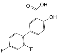
Drug-induced pneumonitis is an uncommon complication of nonsteroidal anti-inflammatory drug administration. Herein is the first reported case of pneumonitis resulting from diflunisal therapy. The patient demonstrated clinical and biopsy evidence of systemic vasculitis. She responded dramatically to administration of systemic glucocorticoids.
Key words: diflunisal; eosinophilic pneumonia; hypersensitivity vasculitis; lung diseases; nonsteroidal anti-inflammatories
Abbreviation: NSAID = nonsteroidal anti-inflammatory drug
Nonsteroidal anti-inflammatory drugs (NSAIDs) have attained popularity in the treatment of a variety of painful and inflammatory conditions. NSAIDs are found in many prescription and over-the-counter preparations. Well-known side effects of these agents include peptic ulcer disease, renal insufficiency, platelet dysfunction, and bronchospasm. Less commonly, NSAIDs can cause pneumonitis. We outline a case in which a patient developed eosinophilic pneumonia with vasculitis following administration of diflunisal (Dolobid).
Case Report
A 21-year-old woman presented to the emergency department at Akron City Hospital on July 27, 1993, with complaints of fever, nausea with emesis, nonproductive cough, pleuritic right-sided chest pain, and painful nodules on her anterior tibial surfaces. She was initially seen at the Akron City Hospital emergency department on July 20 for musculoskeletal neck pain. A WBC count at that time was 9,100/[mm.sup.3] with 2% eosinophils. On July 25, she began receiving diflunisal for her neck pain. Her medical history disclosed that she had not recently traveled and that she had no risk factors for acquisition of HIV. She denied recent use of any other NSAIDs and had taken ibuprofen in the past without adverse effects. Examination on July 27 revealed her to be in no acute distress. Pertinent physical findings included a temperature of 38.0[degrees]C, bibasilar rales, and erythematous, tender, nonblanching nodules on both anterior tibial surfaces. A chest x-ray film revealed bibasilar reticulonodular opacifications. The WBC count was now 12,000/[mm.sup.3] with 19% eosinophils. The hematocrit value was 34.3%. The erythrocyte sedimentation rate was 39 mm/h. Renal and hepatic profiles were essentially normal. Blood, urine, and sputum cultures were obtained, and she was admitted for further evaluation. Diflunisal treatment was stopped and antibiotics were held pending further workup.
When the patient remained febrile and developed purulent sputum, ampicillin-clavulanic acid and erythromycin were begun empirically on day 2. That evening, her respiratory status began to deteriorate and the arterial blood gas level obtained off supplemental oxygenation revealed her [Po.sub.2] to be 58.9 mm Hg. She was placed on supplemental oxygen. The patient's leukocytosis and eosinophilia persisted. She was seen by the pulmonary service on day 4 and was thought to have pulmonary infiltrates with eosinophilia syndrome secondary to NSAID use. A chest x-ray film now showed progressing infiltrates and small effusions (Fig 1). All cultures, including parasite screens, remained negative for bacterial organisms. Her leukocyte count peaked at 29,000/[mm.sup.3] with 44% eosinophils on day 4.
On day 4, she underwent fiberoptic bronchoscopy with BAL and transbronchial biopsy. A BAL fluid sample included an abundance of leukocytes with 53% eosinophils. Other pertinent serologic findings, including angiotensin-converting enzyme level, Legionella antibody titer, mycoplasma IgM, antinuclear antibody, rheumatoid factor, and IgE level, were all within normal limits for our laboratory. Cultures from the BAL sample and of blood failed to grow organisms. The transbrochial biopsy was consistent with eosinophilic pneumonia. There were intra-alveolar and interstitial infiltrates predominantly consisting of eosinophils. Also noted was alveolar lining cell hyperplasia and proteinaceous exudate within alveolar spaces. Blood vessels were affected by endothelial cell hyperplasia and an eosinophilic vasculitis (Fig 2).
By the time of the bronchoscopy, her erythema nodosum had nearly resolved. Oral prednisone, 30 mg three times daily, was initiated on day 6, followed by rapid clinical improvement and decline in eosinophilia. On day 7, she required less supplemental oxygen and her WBC count was 23,000/[mm.sup.3] with 3% eosinophils. A subsequent chest x-ray film revealed interval clearing of the pulmonary infiltrates. On day 8, supplemental oxygen was discontinued and her WBC count was 21,700/[mm.sup.3] with 0% eosinophils. She was discharged home on a tapering dose of prednisone. She returned to the outpatient clinic for follow-up once and at that time was completely asymptomatic. She was told to avoid all NSAIDs and is presently unavailable for follow-up.
Discussion
Diflunisal is a salicylic acid derivative and has been reported to cause such hypersensitivity reactions as interstitial nephritis, necrotizing fasciitis, Stevens-Johnson syndrome, erythema multiforme, and cholestatic jaundice.[1-4] Herein is reported a case of eosinophilic pneumonitis and eosinophilic vasculitis related to the use of diflunisal. This patient is the first reported to have developed pneumonitis consequent to diflunisal ingestion, and this is the first case of eosinophilic vasculitis (represented by lung biopsy findings and erythema nodosum) identified in NSAID-induced pneumonitis. This patient's workup revealed no other apparent cause for her illness, and the temporal relationship to her intake of diflunisal makes it the likely offending agent.
With the assistance of MEDLINE and bibliographic cross-references, we reviewed the world medical literature that pertains to NSAID-induced pneumonitis. We identified nearly 70 cases in which a causal role for an NSAID appeared likely. Many of these cases are contained in prior reviews.[5-7] Using data gleaned from our search, we tabulated the epidemiologic, clinical, and laboratory features of NSAID-induced pneumonitis (data and references available upon request).
Seventeen different NSAIDs have now been reported to induce pneumonitis. In the European literature, azapropazone was the most cited (15 cases). Naproxen was the most frequent cause in the American literature (11 cases). The typical duration of exposure to an NSAID prior to the onset of pulmonary symptoms was 1 to 2 weeks. Most patients were older than 55 years and nearly two thirds of those affected were women. These data may reflect an age and gender predisposition or, more likely, results from greater use of NSAIDs by these groups.
NSAID-induced pneumonitis usually presents as eosinophilic pneumonia. The most common symptoms are fever, cough, dyspnea, and malaise. The most common physical examination findings are fever, bilateral rales, and a generalized rash. Leukocytosis, peripheral eosinophilia, elevated erythrocyte sedimentation rate (sometimes greater than 100 mm/h), and sputum eosinophilia usually are found. Of note, we discovered that liver transaminase levels were elevated in nine cases. Nearly all chest x-ray films demonstrated bilateral interstitial opacifications.
BAL was performed in 13 patients, 8 of whom had eosinophilia. In all 22 patients from whom lung biopsy specimens were obtained, histologic findings were abnormal. The most common findings on biopsy were intra-alveolar and interstitial infiltrates, thickened alveolar walls, interstitial fibrosis, and tissue eosinophilia.
Treatment requires withdrawal of the offending NSAID. Corticosteroids are reserved for the more serious or refractory cases. In the majority of cases in the literature, resolution required only cessation of the NSAID. Those patients receiving corticosteroids responded rapidly. Nineteen patients were either intentionally or inadvertently rechallenged with the same NSAID and all suffered recurrent pneumonitis, generally within 48 h of readministration.
NSAIDs likely cause pneumonitis through a hypersensitivity mechanism. Support for this mechanism includes the development of peripheral, sputum, and tissue eosinophilia; the presence of fever and rash; elevation of liver transaminases perhaps through immune-mediated hepatocellular injury as described in other types of drug hypersensitivities; the interval between administration and clinical onset, typically 2 weeks following initial exposure and then within hours of rechallenge; resolution with corticosteroids; and, as demonstrated in our patient, the presence of systemic vasculitis. A prostaglandin-mediated phenomenon is unlikely as several affected patients had taken other NSAIDs without reaction.
Medications, including NSAIDs, should be part of the differential diagnosis of diffuse pulmonary infiltrates, particularly in those cases associated with eosinophilia or failure to respond to such conventional therapy as antibiotics. A low index of suspicion may result in inappropriate and even dangerous interventions. Fourteen patients in our review had received antibiotics prior to definitive therapy for the NSAID-induced pneumonitis. Treatment in two cases included potentially harmful cytotoxic agents (azathioprine and cyclophosphamide). This syndrome is not always benign. Two patients developed residual pulmonary fibrosis probably due to chronic exposure, and another patient died as a result of acute exposure. When taking a medication history, the physician must include over-the-counter drugs now that a growing number of NSAIDs are becoming available without a prescription. Early diagnosis of acute pneumonitis is important since removal of the offending agent generally leads to rapid resolution. On the other hand, misdiagnosis can lead to prolonged exposure with development of progressive pulmonary fibrosis.
There is another caveat. Interstitial lung disease, whether acute, such as eosinophilic pneumonitis, or chronic, such as interstitial fibrosis, can result from either underlying collagen vascular disorders or the medications used to treat them. For example, consider the possible causes in the hypothetical patient with active rheumatoid arthritis who presents with diffuse pulmonary infiltrates. Potential causes would include rheumatoid lung, infections permitted by immunosuppression related to corticosteroids, or drug-induced lung disease caused by an NSAID or methotrexate.
Lastly, it would seem prudent that a patient who has had this type of reaction to one class of NSAID avoid all other classes. The mechanism for NSAID-induced pneumonitis remains uncertain, and rechallenge with the same drug or cross reaction with another drug may prove dangerous.
References
[1] Cook DJ, Achong MR, Murphy FR. Three cases of diflunisal hypersensitivity. Can Med Assoc J 1988; 138:1029-30
[2] Street ML, Winkelmann RK. Lichenoid photoreactive epidermal necrosis with diflunisal. J Am Acad Dermatol 1989; 20:850-51
[3] Roetzheim RG, Herold AH, VanDurme DJ. Nonpigmenting fixed drug eruption caused by diflunisal. J Am Acad Dermatol 1991; 24:1021-22
[4] Chan LK, Winearls CG, Oliver DO. Acute interstitial nephritis and erythroderma associated with diflunisal. BMJ 1980; 280:84-5
[5] Goodwin SD, Glenny RW. Nonsteroidal anti-inflammatory drug-associated pulmonary infiltrates with eosinophilia. Arch Intern Med 1992; 152:1521-24
[6] Khalil H, Molinary E, Stoller JK. Diclofenac (Voltaren)-induced eosinophilic pneumonitis. Arch Intern Med 1993; 153:1649-52
[7] Pfitzenmeyer P, Meier M, Zuck P, et al. Piroxicam induced pulmonary infiltrates and eosinophilia. J Rheumatol 1994; 21:1573-77
COPYRIGHT 1997 American College of Chest Physicians
COPYRIGHT 2004 Gale Group


