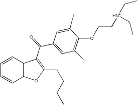
Chemical structure of amiodarone
Find information on thousands of medical conditions and prescription drugs.

Amiodarone

Amiodarone belongs to a class of drugs called Vaughan-Williams Class III antiarrhythmic agent. It is used in the treatment of a wide range of cardiac tachyarhthmias, including both ventricular and supraventricular (atrial) arrhythmias. The chemical name for amiodarone is 2-butyl-3-benzofuranyl 4--3,5-diiodophenyl ketone hydrochloride. more...

Home
Diseases
Medicines
A
8-Hour Bayer
Abacavir
Abamectin
Abarelix
Abciximab
Abelcet
Abilify
Abreva
Acamprosate
Acarbose
Accolate
Accoleit
Accupril
Accurbron
Accure
Accuretic
Accutane
Acebutolol
Aceclidine
Acepromazine
Acesulfame
Acetaminophen
Acetazolamide
Acetohexamide
Acetohexamide
Acetylcholine chloride
Acetylcysteine
Acetyldigitoxin
Aciclovir
Acihexal
Acilac
Aciphex
Acitretin
Actifed
Actigall
Actiq
Actisite
Actonel
Actos
Acular
Acyclovir
Adalat
Adapalene
Adderall
Adefovir
Adrafinil
Adriamycin
Adriamycin
Advicor
Advil
Aerobid
Aerolate
Afrinol
Aggrenox
Agomelatine
Agrylin
Airomir
Alanine
Alavert
Albendazole
Alcaine
Alclometasone
Aldomet
Aldosterone
Alesse
Aleve
Alfenta
Alfentanil
Alfuzosin
Alimta
Alkeran
Alkeran
Allegra
Allopurinol
Alora
Alosetron
Alpidem
Alprazolam
Altace
Alteplase
Alvircept sudotox
Amantadine
Amaryl
Ambien
Ambisome
Amfetamine
Amicar
Amifostine
Amikacin
Amiloride
Amineptine
Aminocaproic acid
Aminoglutethimide
Aminophenazone
Aminophylline
Amiodarone
Amisulpride
Amitraz
Amitriptyline
Amlodipine
Amobarbital
Amohexal
Amoxapine
Amoxicillin
Amoxil
Amphetamine
Amphotec
Amphotericin B
Ampicillin
Anafranil
Anagrelide
Anakinra
Anaprox
Anastrozole
Ancef
Android
Anexsia
Aniracetam
Antabuse
Antitussive
Antivert
Apidra
Apresoline
Aquaphyllin
Aquaphyllin
Aranesp
Aranesp
Arava
Arestin
Arestin
Argatroban
Argatroban
Argatroban
Argatroban
Arginine
Arginine
Aricept
Aricept
Arimidex
Arimidex
Aripiprazole
Aripiprazole
Arixtra
Arixtra
Artane
Artane
Artemether
Artemether
Artemisinin
Artemisinin
Artesunate
Artesunate
Arthrotec
Arthrotec
Asacol
Ascorbic acid
Asmalix
Aspartame
Aspartic acid
Aspirin
Astemizole
Atacand
Atarax
Atehexal
Atenolol
Ativan
Atorvastatin
Atosiban
Atovaquone
Atridox
Atropine
Atrovent
Augmentin
Aureomycin
Avandia
Avapro
Avinza
Avizafone
Avobenzone
Avodart
Axid
Axotal
Azacitidine
Azahexal
Azathioprine
Azelaic acid
Azimilide
Azithromycin
Azlocillin
Azmacort
Aztreonam
B
C
D
E
F
G
H
I
J
K
L
M
N
O
P
Q
R
S
T
U
V
W
X
Y
Z

History

Amiodarone was initially developed in 1961 in Belgium as a treatment for angina. It was widely used throughout Europe as an anti-anginal medication, and was soon found to suppress arrhythmias.

Dr. Bramah Singh determined that amiodarone and sotalol belonged to a new class of antiarrhythmic agents (what would become the class III antiarrhythmic agents) that would prolong repolarization of the cardiac action potential. Based on this, the Argentinian physician Dr. Mauricio Rosenbaum began using amiodarone to treat his patients who suffered from supraventricular and ventricular arrhythmias, with impressive results. Based on papers written by Dr. Rosenbaum, physicians in the United States began prescribing amiodarone to their patients with potentially life-threatening arrhythmias in the late 1970s. By that time, amiodarone was commonly prescribed throughout Europe for the treatment of arrhythmias. Because amiodarone was not approved by the FDA for use in the United States at the time, physicians were forced to directly obtain amiodarone from pharmaceutical companies in Canada and Europe.

The FDA was reluctant to officially approve the use of amiodarone, since initial reports had shown increased incidence of serious pulmonary side-effects of the drug. In the mid 1980s, the European pharmaceutical companies began putting pressure on the FDA to approve amiodarone by threatening to cut the supply to the American physicians if it was not approved. In December of 1985, amiodarone was approved by the United States FDA for the treatment of arrhythmias. This makes amiodarone one of the few drugs approved by the FDA without rigorous randomized clinical trials.

Dosing

Amiodarone is available in oral and intravenous formulations. Orally, it is available under the trade names Pacerone® (produced by Upsher-Smith Laboratories, Inc.) and Cordarone® (produced by Wyeth-Ayerst Laboratories) in 200 mg and 400 mg tablets. It is also available in intravenous ampules and vials, typically in 150mg increments.

The dose of amiodarone administered is tailored to the individual and the dysrhythmia that is being treated. When administered orally, the bioavailability of amiodarone is quite variable. Absorption ranges from 22 to 95%, with better absorption when it is given with food.

Amiodarone is fat-soluble, and tends to concentrate in tissues including fat, muscle, liver, lungs, and skin. This confers a high volume of distribution (5000 liters in a 70kg adult) and a long half-life. Due to the long half-life of amiodarone, oral loading typically takes days to weeks.

Read more at Wikipedia.org


[List your site here Free!]


Amiodaronoma: one of multiple forms of amiodarone pulmonary toxicity
From CHEST, 10/1/05 by Julie Jarand

INTRODUCTION: Amiodarone pulmonary toxicity most commonly takes the form of interstitial pneumontitis (1). Here we report a case of amiodarone-induced lung injury presenting as a pulmonary mass.

CASE PRESENTATION: A 66 year old woman presented to hospital with dyspnea secondary to congestive heart failure and high grade atrioventricular block. She underwent insertion of a biventricular pacemaker without complication. A routine post-procedure chest radiograph revealed a right upper lobe mass. She was referred for further pulmonary assessment. She has a 50 pack year smoking history. Her past medical history is significant for severe left ventricular dysfunction, supraventricular tachycardia (SVT), recently diagnosed hypothyroidism, hyperlipidemia and remote pulmonary embolism. SVT had been treated with amiodarone 200mg daily for four years (total cumulative dose approximately 300g). Her dyspnea had resolved following pacemaker insertion and medical management of heart failure. She denied any other respiratory or constitutional symptoms. Physical examination was unremarkable. ALT was mildly elevated (67 U/L). A nonenhanced chest computed tomography (CT) scan showed an irregular right upper lobe mass measuring 3.0 x 2.3 cm (Figure 1). A smaller (1.8 x 1.2 cm) right lower lobe peripheral wedge shaped lesion was also noted. There was increased density of both lesions (80 Hounsfield units) as well as hyperdensity of the liver. Percutaneous fine needle aspirate of the right upper lobe mass was performed. Pathology revealed chronic interstitial inflammation, organizing pneumonia, intra-alveolar aggregates of foamy macrophages and type 2 pneumocyte hyperplasia. There was no evidence of malignancy. These results are consistent with pulmonary changes due to amiodarone therapy (1). Repeat imaging three months after discontinuation of amiodarone showed complete resolution of radiologic abnormalities (Figure 2).

[FIGURE OMITTED]

DISCUSSIONS: This case represents an unusual presentation of amiodarone pulmonary toxicity. There have been isolated case reports of similar presentations over the last 20 years, but none of these have correlated radiographic and CT imaging with light and electron microscopy (24). Previous reports have postulated that these solitary depositions are due to increased localized accumulation of the drug in an area of previous inflammation (5). Interestingly, all previous reported cases describe masses in the upper lobes, particularly right upper lobe. Our case highlights the fact that primary lung cancer needs to be excluded in a long standing smoker with a lung mass. Also, one needs to consider amiodarone toxicity as a potential, usually reversible, etiology. With an increasing population at risk for both lung cancer and cardiovascular disease, the need for this distinction will become increasingly important. It also demonstrates that amiodarone toxicity can occur alter many years of low dose therapy. Previous studies support that toxicity correlates more closely with total cumulative dose and duration than with serum drug levels (1). This case is in keeping with previous reports who had concomitant thyroid disease and/or liver toxicity. Abnormalities in other organ systems should prompt physicians to look for pulmonary amiodarone toxicity in asymptomatic patients. Diagnosis can be made by combination of clinical, radiologic and/or pathologic findings with improvement after discontinuation of the drug.

CONCLUSION: Amiodarone-induced toxicity should be included in the differential diagnosis of pulmonary mass lesions in patients receiving amiodarone.

REFERENCES:

(1) Camus P et al. Amiodarone pulmonary toxicity. Clin Chest Med 2004;25:65-75.

(2) Arnon R et al. Amiodarone pulmonary toxicity presenting as a solitary lung mass. Chest 1988;93:425-427.

(3) Piccione W Jr et al. Amiodarone-induced pulmonary mass. Ann Thorac Surg 1989;47:918-919.

(4) Rodriguez-Garcia JL et al. Pulmonary mass and multiple pulmonary nodules mimicking a lung neoplasm as amiodarone-induced pulmonary toxicity. Eur J Intern Med 2001;12:372-376.

(5) Kuhlman JE et al. Amiodarone Pulmonary Toxicity: CT findings in symptomatic patients. Radiol 1990;177:121-125.

DISCLOSURE: Julie Jarand, None.

Julie Jarand MD * Jessi Minion MD Andrew Lee MD Francis Green MD Richard Leigh MD University of Calgary, Calgary, AB, Canada

COPYRIGHT 2005 American College of Chest Physicians
COPYRIGHT 2005 Gale Group

Return to Amiodarone
Home Contact Resources Exchange Links ebay