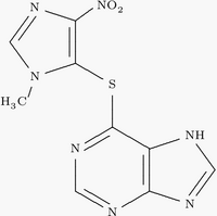
Structure formula of azathioprine
Find information on thousands of medical conditions and prescription drugs.

Azathioprine

Azathioprine is a chemotherapy drug, now rarely used for chemotherapy but more for immunosuppression in organ transplantation and autoimmune disease such as rheumatoid arthritis or inflammatory bowel disease or Crohn's disease. It is a pro-drug, converted in the body to the active metabolite 6-mercaptopurine. more...

Home
Diseases
Medicines
A
8-Hour Bayer
Abacavir
Abamectin
Abarelix
Abciximab
Abelcet
Abilify
Abreva
Acamprosate
Acarbose
Accolate
Accoleit
Accupril
Accurbron
Accure
Accuretic
Accutane
Acebutolol
Aceclidine
Acepromazine
Acesulfame
Acetaminophen
Acetazolamide
Acetohexamide
Acetohexamide
Acetylcholine chloride
Acetylcysteine
Acetyldigitoxin
Aciclovir
Acihexal
Acilac
Aciphex
Acitretin
Actifed
Actigall
Actiq
Actisite
Actonel
Actos
Acular
Acyclovir
Adalat
Adapalene
Adderall
Adefovir
Adrafinil
Adriamycin
Adriamycin
Advicor
Advil
Aerobid
Aerolate
Afrinol
Aggrenox
Agomelatine
Agrylin
Airomir
Alanine
Alavert
Albendazole
Alcaine
Alclometasone
Aldomet
Aldosterone
Alesse
Aleve
Alfenta
Alfentanil
Alfuzosin
Alimta
Alkeran
Alkeran
Allegra
Allopurinol
Alora
Alosetron
Alpidem
Alprazolam
Altace
Alteplase
Alvircept sudotox
Amantadine
Amaryl
Ambien
Ambisome
Amfetamine
Amicar
Amifostine
Amikacin
Amiloride
Amineptine
Aminocaproic acid
Aminoglutethimide
Aminophenazone
Aminophylline
Amiodarone
Amisulpride
Amitraz
Amitriptyline
Amlodipine
Amobarbital
Amohexal
Amoxapine
Amoxicillin
Amoxil
Amphetamine
Amphotec
Amphotericin B
Ampicillin
Anafranil
Anagrelide
Anakinra
Anaprox
Anastrozole
Ancef
Android
Anexsia
Aniracetam
Antabuse
Antitussive
Antivert
Apidra
Apresoline
Aquaphyllin
Aquaphyllin
Aranesp
Aranesp
Arava
Arestin
Arestin
Argatroban
Argatroban
Argatroban
Argatroban
Arginine
Arginine
Aricept
Aricept
Arimidex
Arimidex
Aripiprazole
Aripiprazole
Arixtra
Arixtra
Artane
Artane
Artemether
Artemether
Artemisinin
Artemisinin
Artesunate
Artesunate
Arthrotec
Arthrotec
Asacol
Ascorbic acid
Asmalix
Aspartame
Aspartic acid
Aspirin
Astemizole
Atacand
Atarax
Atehexal
Atenolol
Ativan
Atorvastatin
Atosiban
Atovaquone
Atridox
Atropine
Atrovent
Augmentin
Aureomycin
Avandia
Avapro
Avinza
Avizafone
Avobenzone
Avodart
Axid
Axotal
Azacitidine
Azahexal
Azathioprine
Azelaic acid
Azimilide
Azithromycin
Azlocillin
Azmacort
Aztreonam
B
C
D
E
F
G
H
I
J
K
L
M
N
O
P
Q
R
S
T
U
V
W
X
Y
Z

Azathioprine was first introduced into clinical practice by Sir Roy Calne, the British pioneer in transplantation. Following the work done by Sir Peter Medawar in discovering the immunological basis of rejection of transplanted tissues and organs, Calne introduced 6-mercaptopurine as an experimental immunosuppressant for kidney transplants. When azathioprine was discovered, he then introduced it as a less toxic replacement for 6-mercaptopurine. For many years, dual therapy with azathioprine and steroids was the standard anti-rejection regime, until cyclosporine was introduced into clinical practice (also by Calne) in 1978.

Azathioprine acts to inhibit purine synthesis necessary for the proliferation of cells, especially leukocytes and lymphocytes. It is a safe and effective drug used alone in certain autoimmune diseases, or in combination with other immunosuppressants in organ transplantation. Its most severe side effect is bone marrow suppression, and it should not be given in conjunction with purine analogues such as allopurinol. The enzyme thiopurine S-methyltransferase (TPMT) deactivates 6-mercaptopurine. Genetic polymorphisms of TPMT can lead to excessive drug toxicity, thus assay of serum TPMT may be useful to prevent this complication.

Mycophenolate mofetil is increasingly being used in place of azathioprine in organ transplantation, but azathioprine certainly still has a major role.

Mycophenolate mofetil (MMF): seven RCTs compared MMF to azathioprine (AZA). MMF reduced the incidence of acute rejection. There was no significant difference in patient survival or graft loss at 1-year or 3-year follow-up. There appeared to be differences in the side-effect profiles of MMF and AZA. No RCTs comparing MMF with azathioprine were identified. Woodroffe R, Yao GL, Meads C, Bayliss S, Ready A, Raftery J, et al. Clinical and cost-effectiveness of newer immunosuppressive regimens in renal transplantation: a systematic review and modelling study. Health Technol Assess 2005;9(21).

Reference

  • Konstantopoulou M, Belgi A, Griffiths KD, Seale JR, Macfarlane AW. Azathioprine-induced pancytopenia in a patient with pompholyx and deficiency of erythrocyte thiopurine methyltransferase. BMJ 2005;330:350-1. PMID 15705694.

Read more at Wikipedia.org


[List your site here Free!]


Anti-CD20 monoclonal antibody in the treatment of pemphigus
From Journal of Drugs in Dermatology, 11/1/05

Anti-CD20 Monoclonal Antibody (Rituximab) in the Treatment of Pemphigus

Arin MJ, et al. Br J Dermatol. 2005;153:620-625.

Summary

The authors present a series of 5 patients with recalcitrant pemphigus, treated with anti-CD20 monoclonal antibody (rituximab), to assess efficacy of this drug in this patient population. Four patients with pemphigus vulgaris and 1 patient with pemphigus foliaceous were enrolled. All patients were stated to have recalcitrant disease, defined as having active lesions despite treatment with the equivalent of greater than 10 mg of prednisolone daily and azathioprine 2 to 2.5 mg/kg daily, or immunosuppression that was equivalent. Patients were given intravenous infusions of 375 mg/[m.sup.2] of rituximab once a week for 4 weeks. Assessments occurred at weekly intervals for the first month, every 2 weeks for the second month, and then monthly. At each assessment, the patients' skin and mucous membranes were examined. In addition, several blood tests were serially drawn including antibody titers to desmoglein 1 and 3, indirect immunofluorescence, and peripheral blood B cell count. The latter was determined by using flow cytometry to evaluate the number of CD19+ B cells. The authors state that estimate of CD20 is "impaired" when rituximab is circulating in the plasma so they looked at CD19+ B cells, as CD19 is a pan-B cell marker. Primary outcome measures included resolution of mucocutaneous lesions, a decrease in the corticosteroid dose to below 7.5 mg of prednisolone daily, and the length of remission. Complete response was defined as no clinically active disease for at least 8 weeks within 1 year after treatment with rituximab. Partial response included patients with a reduction in active lesions by at least 50% of the initial body area of involvement. Minimal response included patients with a reduction in active lesions by less than 50%. Of the 5 patients treated with rituximab, 3 were complete responders and 2 were partial responders. Of the 3 complete responders, I had no evidence of clinical disease at 18 months post-treatment and required no supplemental medication. Another had no disease at 24 months post-treatment, requiring only 500 mg of mycophenolate mofetil daily. The third had no disease at 36 months post-treatment, needing only 1 mg of prednisolone daily. The partial responders were classified as having "minor disease flares" during a 10-month observation period after rituximab treatment. However, both patients were able to decrease the dosages of immunosuppressive therapy post-treatment compared with treatment at the time of rituximab infusion. Through serial monitoring of CD19+ B cell counts, a correlation was noted between improvement of disease and a decrease in the number of these cells. No serious adverse events were noted in any of the treated patients.

Comment

The authors present a series of patients with recalcitrant pemphigus who were treated with rituximab. Given the rarity of these diseases, the sample size is relatively adequate, though it is important to note that one of the patients had previously been reported in an article published in the British Journal of Dermatology in 2003. The results are difficult to interpret from the data presented in this article. Information regarding the treatment regimen of each patient is listed in a chart within the article, indicating regimens only at the time of rituximab infusion and then "current treatment." It does not delineate the time point after rituximab infusion that immunosuppressive treatment was reduced. In addition, the reader must speculate as to the treatment regimen prior to rituximab infusion. Also, time of response to treatment is only addressed graphically and graph curves for some of the patients seem to already be decreasing prior to rituximab infusion. As rituximab does not appear to have a quick onset of action and since patients were also on concomitant immunosuppressive therapy, it is difficult to assess what role if any rituximab had in the remission of lesions in these patients. Perhaps a longer course of immunosuppressive therapy was more important in clearing lesions than rituximab. Lastly, the criteria used for refractory disease in this article is by no means standardized, considering the lower limit of daily corticosteroid use was required to be only 10 mg for patients to be labeled as refractory. Rituximab may be of benefit in the treatment of pemphigus, but it is difficult to state this from how the data is presented in this article. Further studies are needed to evaluate its efficacy, clearly delineating pretreatment regimens and history of immunosuppressive therapy, changes in treatment regimens, and the time course of these changes.

COPYRIGHT 2005 Journal of Drugs in Dermatology, Inc.
COPYRIGHT 2005 Gale Group

Return to Azathioprine
Home Contact Resources Exchange Links ebay